girdle of 21–22 longitudinal rows of cilia in mid-body
oral opening in mid-body
anterior and posterior end naked
contractile vacuole subterminal
one caudal cilium in an eccentric position
fast swimming, restless, jerkily dancing
Urozona buetschlii
Urozona buetschlii is a very difficult object for microphotography. The ciliate is not only very small, but also an extremely fast swimmer. In samples from sludge, one often finds many specimens jumping between detritus flakes at high speed. In live observation, the shape and structure of Urozona buetschlii cannot be recognized. If one reduces the amount of water under the coverslip to slow down movements, the specimens denature immediately. So you have to try to photograph specimens which swim for fractions of a second near the coverslip. In my experience, this only works with a high reject rate of failed photos, but eventually leads to success. This is how the photos shown below were taken.
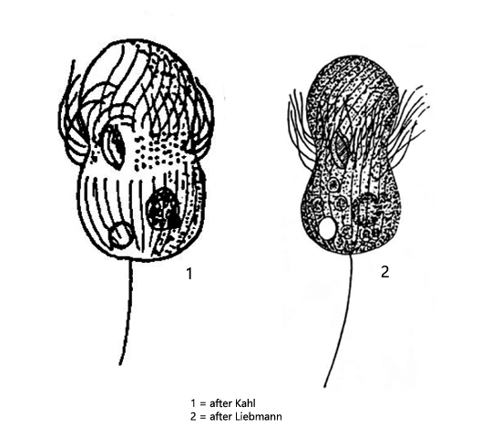
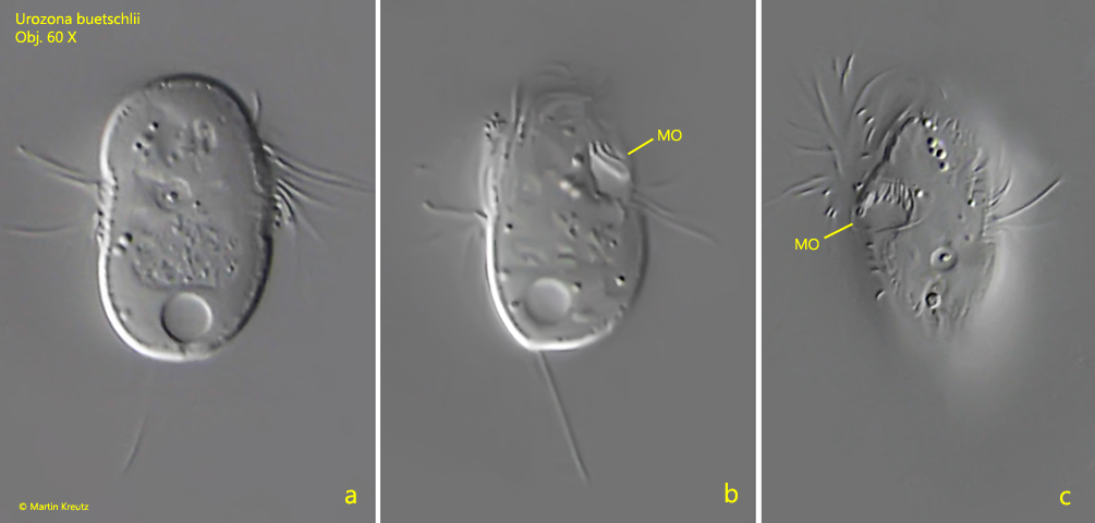
On the flashed photos you can clearly see the typical dumbbell shape of Urozona buetschlii (s. figs. 1 a and 2 a-c). A belt of cilia runs around the tapered mid region, which provides all the propulsion (s. fig. 2 a). The mouth opening is at the level of this belt of cilia, in a short gap (s. figs. 1 b and 1 c). The terminal caudal cilium is located eccentrically (s. fig. 2a). All these details can be seen only in flashed microphotographs. I suspect that the earlier authors, who did not have this technique yet available, made their drawing from fixed material.
Fig. 1 a-c:Urozona buetschlii. L = 25 µm. A freely swimming specimen. MO = mouth opening. Obj. 60 X.
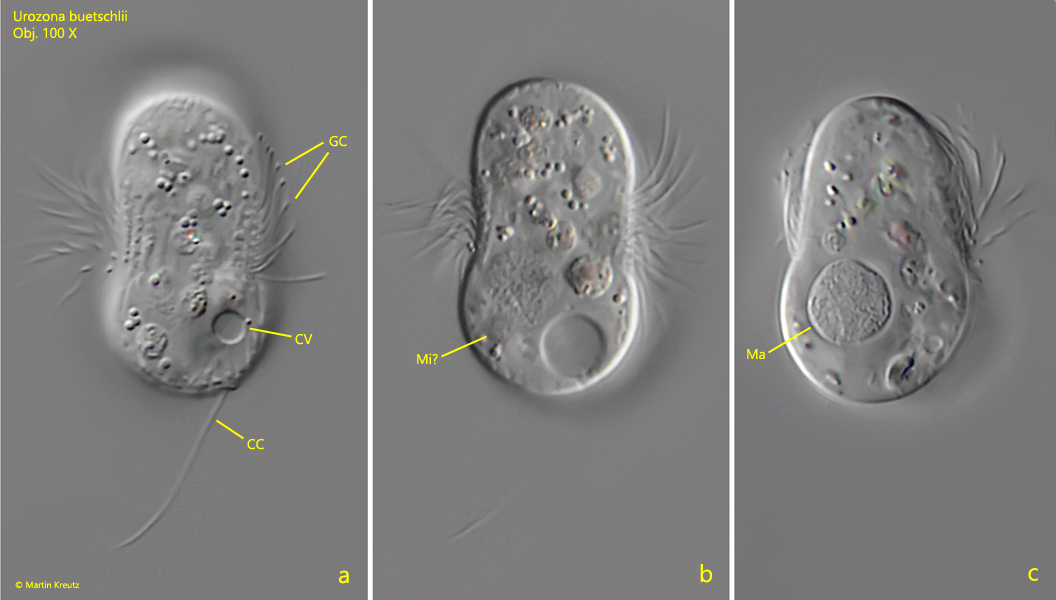
Fig. 2 a-c:Urozona buetschlii. L = 27 µm. A second freely swimming specimen. Note the girdle of cilia (GC) in mid-body and the caudal cilium (CC) in eccentric position. CV = contractile vacuole, Ma = macronucleus, Mi? = likely the micronucleus. Obj. 100 X.
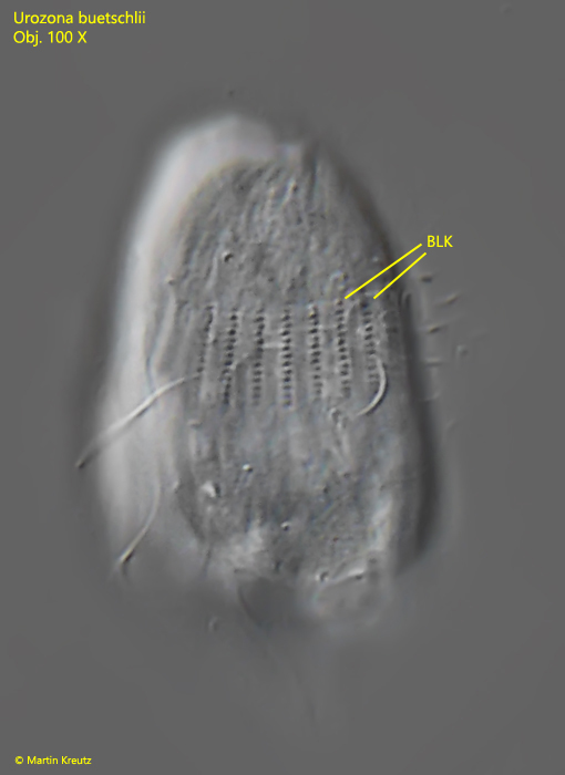
Fig. 3:Urozona buetschlii. The basal bodies of the longitudinal kineties (BLK) in a squashed specimen. Obj. 100 X.
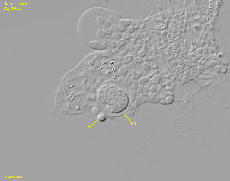
Fig. 4:Urozona buetschlii. The spherical macronucleus (Ma) and the adjacent micronucleus (Mi) in a strongly squashed specimen. Obj. 100 X.