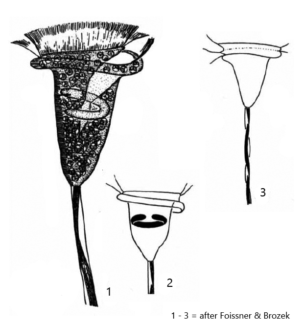
contracted cells pyriform with bulged anterior region
stalk 300 – 500 µm long, contracts in spirals
plasm green due to symbiotic algae
symbiotic algae 5 – 6 µm in diameter, spherical, one pyrenoid
one contracile vacuole
macronucleus horseshoe-shaped in anterior half
pellicle with very fine transverse striation (in average 107 lines)
solitary or in pseudocolonies
Vorticella chlorostigma
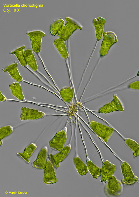
I found Vorticella chlorostigma for the first time in June 1998 in Simmelried. Afterwards again in 2021 and 2022. The species seems to occur mainly in the warm summer months. It is very easy to recognize by the broad, umbrella-shaped peristome and by the green coloration due to the symbiotic algae. The cells are about 100 µm in length and are also quite conspicuous because of their size. In my population cells with up to 120 µm length were present. In 1996 Foissner and Brozek published a detailed re-description of the species (s. Literature). Their re-description fully matches the characteristics of the indivuduals in my population.
Fig. 1: Vorticella chlorostigma. L = 97 – 121 µm. A pseudocolony of about 30 individuals. Obj. 10 X.
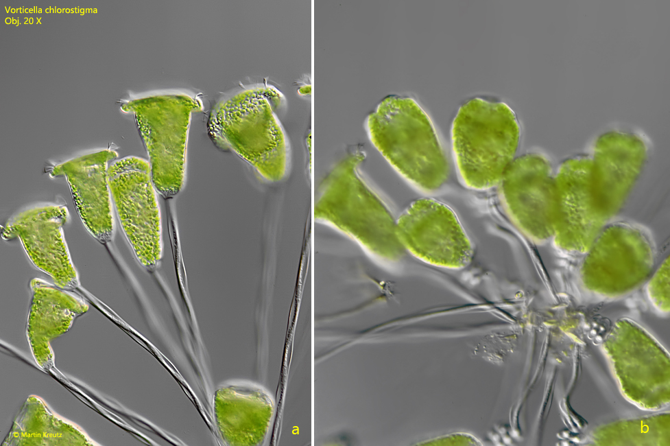
Fig. 2a-b: Vorticella chlorostigma. L = 97 – 121 µm. Fully extended individuals (a) and contracted individuals (b) in a pseudocolony. Obj. 20 X.
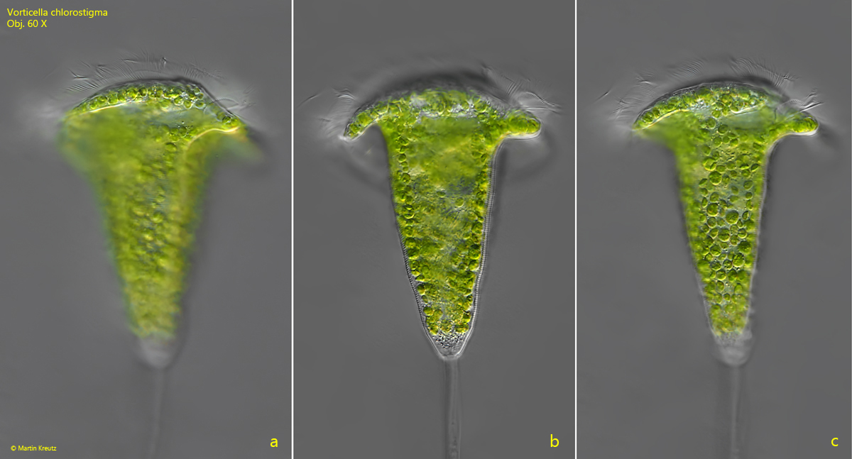
Fig. 3 a-c: Vorticella chlorostigma. L = 121 µm. A fully extended specimen in three focal planes. Obj. 60 X.
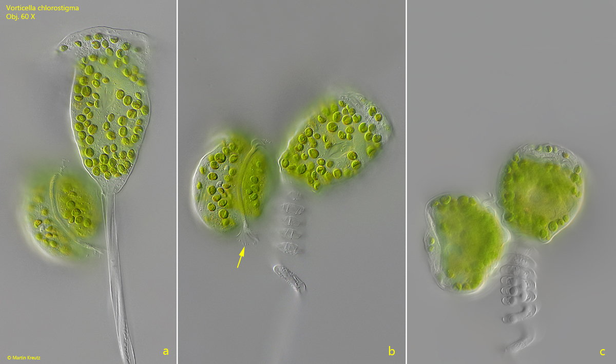
Fig. 4 a-c: Vorticella chlorostigma. L = 98 µm. Different states from an fully extended to a contracted specimen. The left cell is a daughter cell after a cell division. The daughter cell is forming a ciliary ring (b, arrow) in the region of the aboral ciliary wreath and will detach from the stalk later. Obj. 60 X.
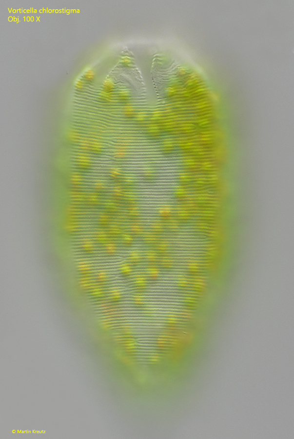
Fig. 5: Vorticella chlorostigma. The fine and delicate transverse striation of the pellicle in a slightly squashed specimen. The distance beween the stripes is 0.62 µm. Obj. 100 X.
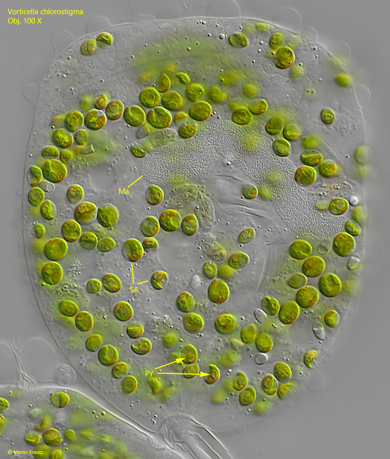
Fig. 6: Vorticella chlorostigma. The horseshoe-shaped macronucleus (Ma) lies transversely in the upper half of the cell. Here it can be seen in a strongly squashed cell. The symbiontic algae (SA) have a diameter of 5-6 µm and possess a distinct pyrenoid (PY). Obj. 100 X.