The peritrichous ciliate Vorticella convallaria is very common and found in practically all of my sampling sites. Often pseudocolonies grow on the vessel wall or on the surface in old samples. The observation of such colonies is usually difficult, because the colonies must be detached, whereby they are often destroyed and the cells contract. Observation is much easier when the pseudocolonies settle on floating coverslips. These pseudocolonies (i.e. single individuals standing in groups) I find very often on floating coverslips. Vorticella convallaria is very easy to recognize. They are always single individuals with a spirally contracting stalk. The cells are usually over 50 µm long, with a J-shaped macronucleus and only one contractile vacuole. In addition, the bell-shaped body is finely striated (in my population the stripe spacing was 0.53 µm), allowing a reliable distinction from Pseudovorticella, where the pellicle is covered with small blisters. In my population the individuals were 50–80 µm long.
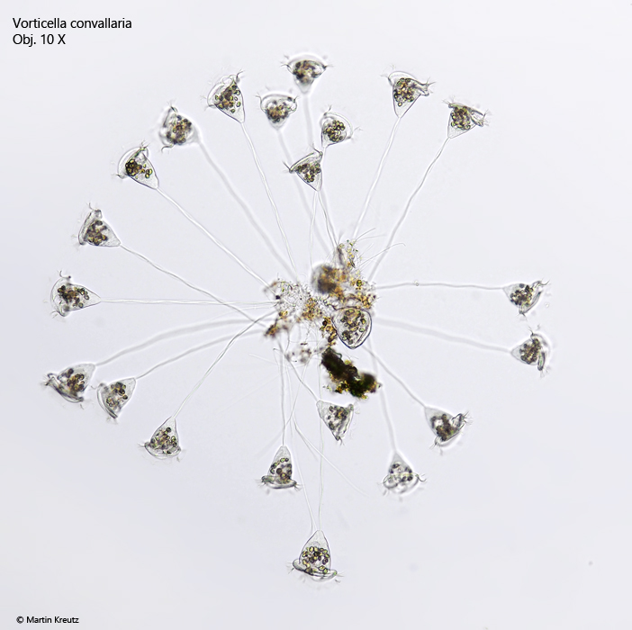
Fig. 1: Vorticella convallaria. A pseudocolony attached to a detritus flake in brightfield illumination. Obj. 10 X.
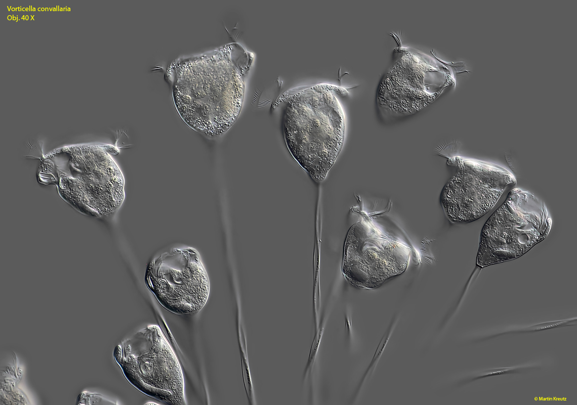
Fig. 2: Vorticella convallaria. Freely moving specimens in a pseudocolony. Obj. 40 X.
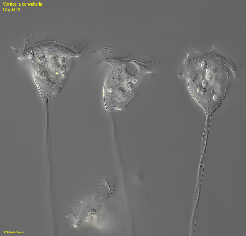
Fig. 3: Vorticella convallaria. L = 58–70 µm. Three extended specimens. The stalks have a length of 130–260 µm. Obj. 60 X.
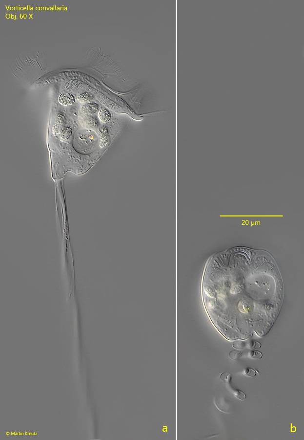
Fig. 4 a-b: Vorticella convallaria. A fully extended specimen with a length of 66 µm (a) and the same specimen fully contracted with a length of 51 µm (b). Obj. 60 X.
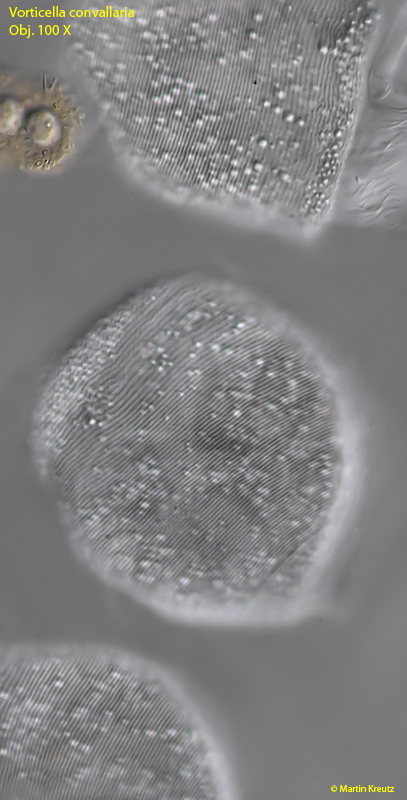
Fig. 5: Vorticella convallaria. In a squashed specimen the fine striation of the pellicle becomes visible. Obj. 100 X.
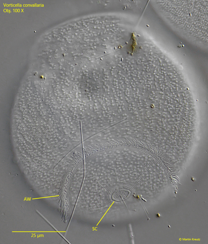
Fig. 6: Vorticella convallaria. A view of the posterior third of a squashed specimen shows the aboral ciliary wreath (AW) as well as the scopula (SC), an organelle for the formation of the stalk. Obj. 100 X.
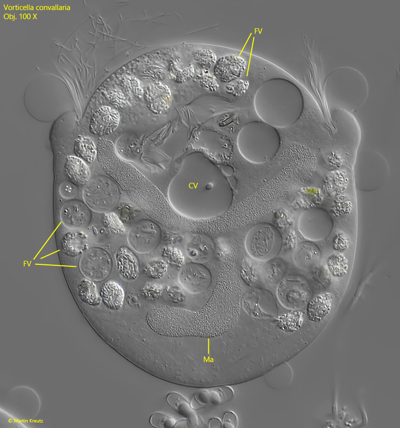
Fig. 7: Vorticella convallaria. A strongly squashed specimen. In the center the contracile vacuole (CV). The J-shaped macronucleus (Ma) is folded in the cell. The food vacuoles (FV) are filled with bacteria in diferent states of digestion process. Obj. 100 X.
In many individuals of the pseudocolonies, I noticed highly refractive bodies in the posterior part of the cells, near the origin of the stalk. In squashed cells I recognized that it was with high probability an infestation by a parasitic fungus. However, I am not entirely sure because the cells were quite small with only 4.8 µm length and I also could not see a nucleus. Possibly the invaders were also bacteria or representatives of the Apicomplexa.
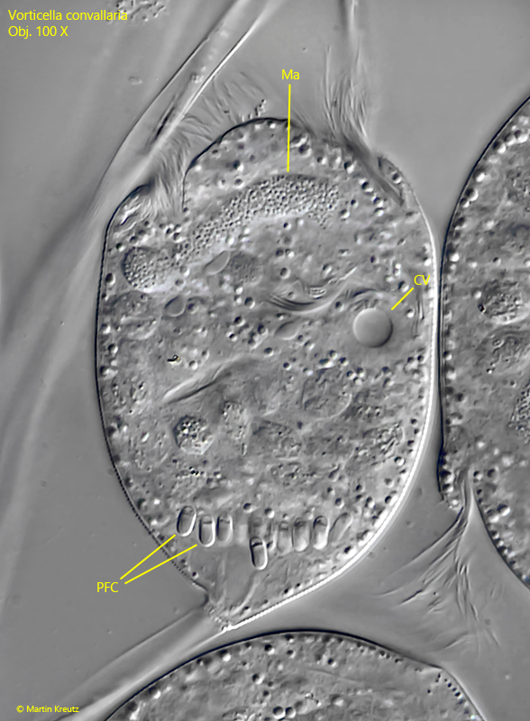
Fig. 8: Vorticella convallaria. Slightly squashed specimen with an accumulation of 4– 5 µm long cells at the stalk base, which may be parasitic fungal cells (PFC). Note the single contractile vacuole (CV). Ma = macronucleus. Obj. 100 X.
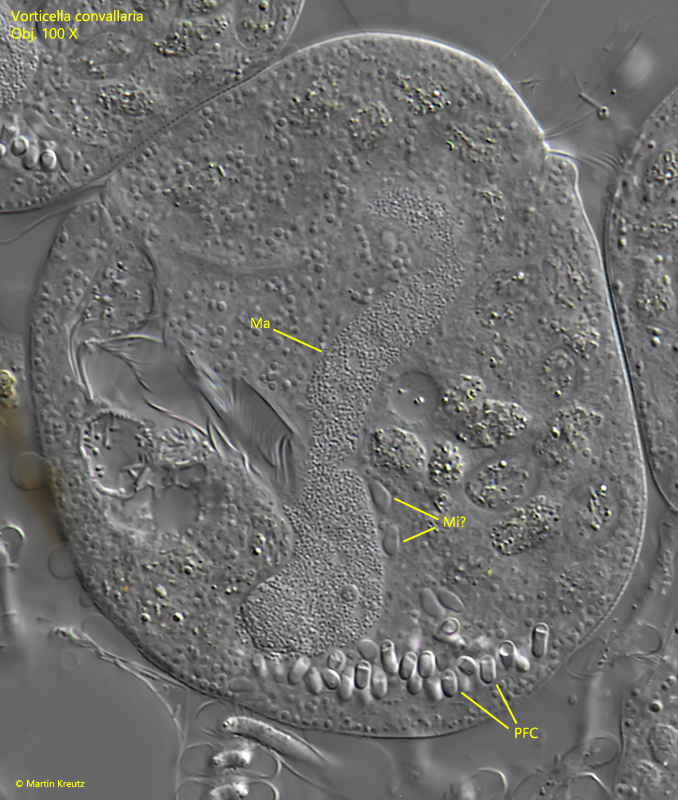
Fig. 9: Vorticella convallaria. A more strongly squashed specimen, also with an accumulation of parasitic fungal cells (PFC). Ma = macronucleus, Mi? = likely the micronuclei which are attached to the J-shaped macronucleus. Obj. 100 X.
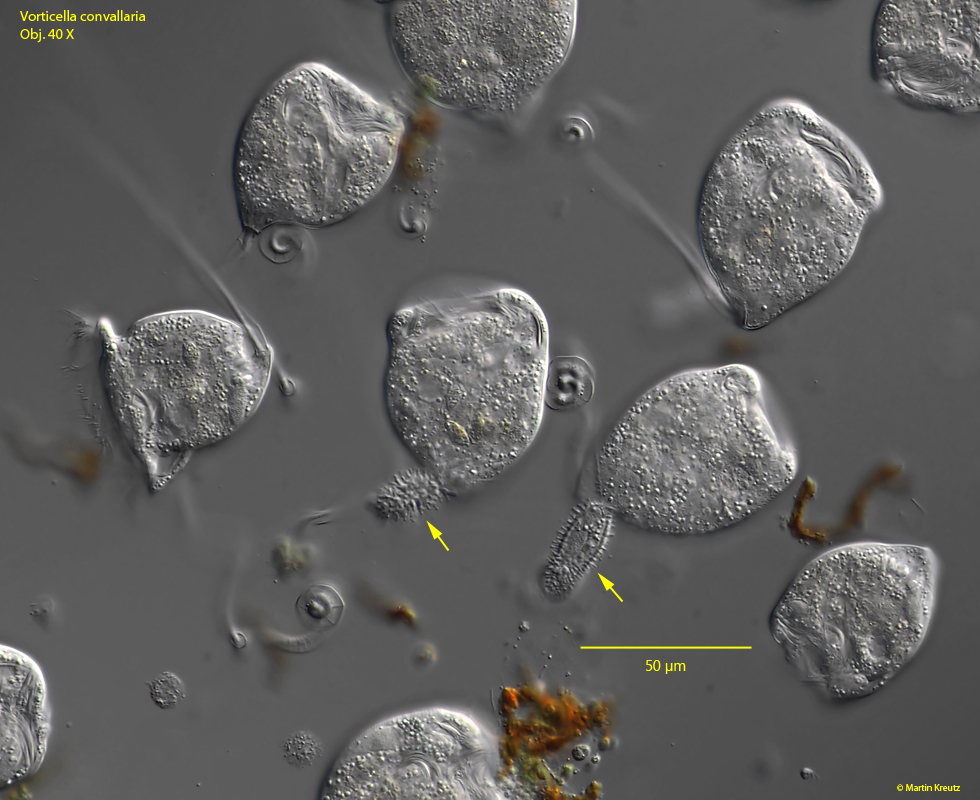
In the pseudocolonies of Vorticella convallaria I examined, I also found more frequent stages of sexual reproduction. This occurs in sessile peritrichous ciliates via the formation of microgametes, which are mobile and attach to the stalk base of sessile zooids. After formation of a cytoplasmic bridge between the microgametes and the sessile zooid, the plasma and genetic material of the microgametes are transferred to the zooid, which is then called a macrogamete. In this process, the microgamete continues to shrink and assumes a shriveled form with a pointed end. I could often find this stage of conjugation.
Fig. 10: Vorticella convallaria. Overview of a pseudocolony with some zooids in conjugation with microgamets (arrows). Obj. 40 X.
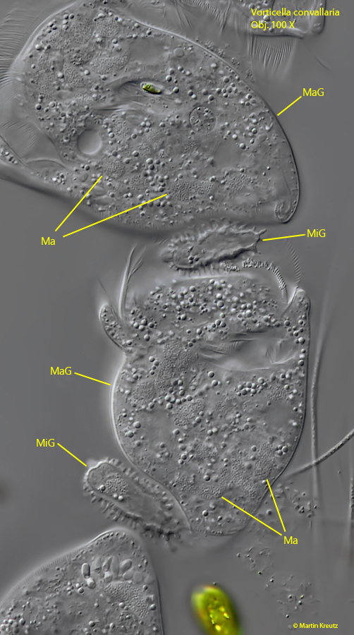
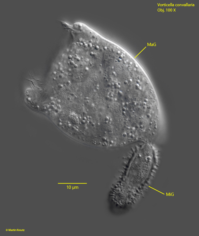
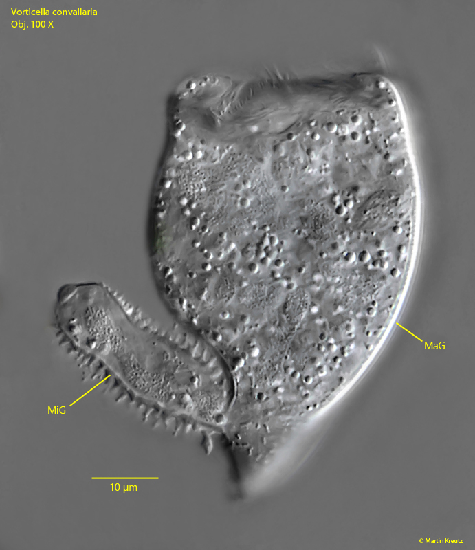
Fig. 11: Vorticella convallaria. During the conjugation the genetic material of the microgamete (MiG) is transferred into the macrogamete (MaG). In this stage the macronucleus (Ma) of the macrogamete begins to disintegrate. Obj. 100 X.
Fig. 12, 13: Vorticella convallaria. Two macrogametes (MaG) in conjugation with microgametes (MiG). Note the thin bridge of cytoplasm between the micro- and macrogamete for the transfer of protoplasm and genetic material (the micronucleus after a meiotic division). Obj. 100 X.
Given the very few morphological differences, several species have been grouped under Vorticella convallaria, such as Vorticella convallaria var. citrina, Vorticella citrina or Vorticella similis (s. “Synonyms” above). Therefore, Vorticella convallaria is also called Vorticella convallariacomplex.
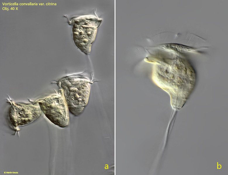
The color of the plasm of Vorticella convallaria can often become distinctly yellow or even lemon yellow. This form was formerly called Vorticella convallaria var. citrina (s. fig. 11 a-b) or Vorticella citrina.
Fig. 14 a-b: Vorticella convallaria var. citrina. A yellow colored variant of Vorticella convallaria. Obj. 40 X.