colonies free-floating, outline ellipsoidal, globular or somewhat irregular
diameter of colonies 50–180 µm
colonies in a distinct gelatinous envelope
cells obvoid or ellipsoidal, 3.5–5 µm x 7–11 µm
cyptoplasm with scattered, small gas vacuoles
cells on distal end of gelatinous stalks, connected in the center (hard to see)
planktonic lifestyle
Woronichinia naegeliana
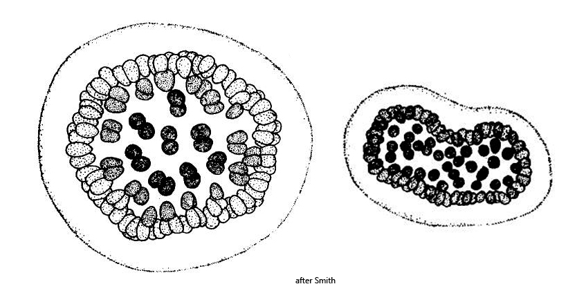
The cyanobacterium Wolonichinia naegelinana was first described as Coelosphaerium naegelianum. After Woronichin was able to detect gelatinous stalks in the colonies after staining with methylene blue, the species was transferred to the genus Wolonichinia. However, the gelatinous stalks are difficult to recognize. I was also unable to detect them in the DIC (s. fig. 2 a-b). The cells of Wolonichinia naegelinana are very large with finely distributed gas vacuoles. In my population, the cells were 4–5 µm x 5–9 µm in size, which clearly distinguishes them from the similar species Coelosphaerium kuetzingianum, whose almost spherical cells are only 2–4 µm in size. Colonies of Wolonichinia and Coelosphaerium can also be confused with Microcystis. In Microcystis, however, the cells are scattered in the gelatinous mass and do not form a hollow sphere.
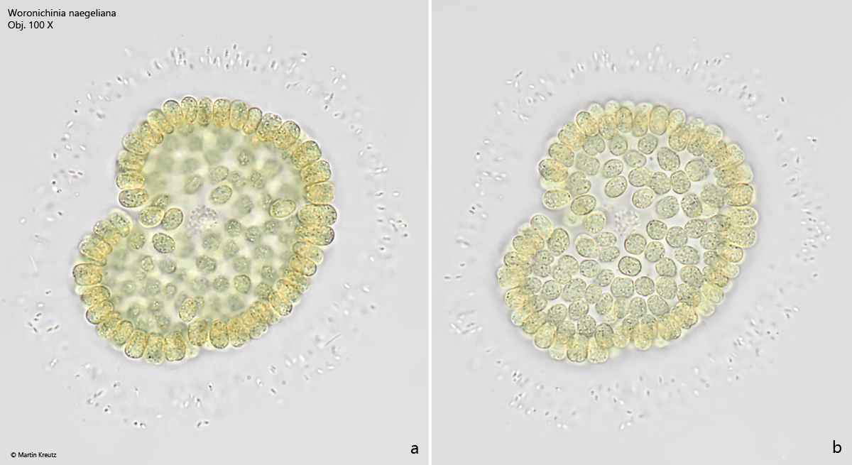
Fig. 1 a-b:Woronichinia naegeliana. D = 54 µm (of colony). Two focal planes of colony in brightfield illumination. The cells arranged in a single layer have a length of 7.1–8.4 µm and a width of 4.0–5.3 µm. Obj. 100 X.
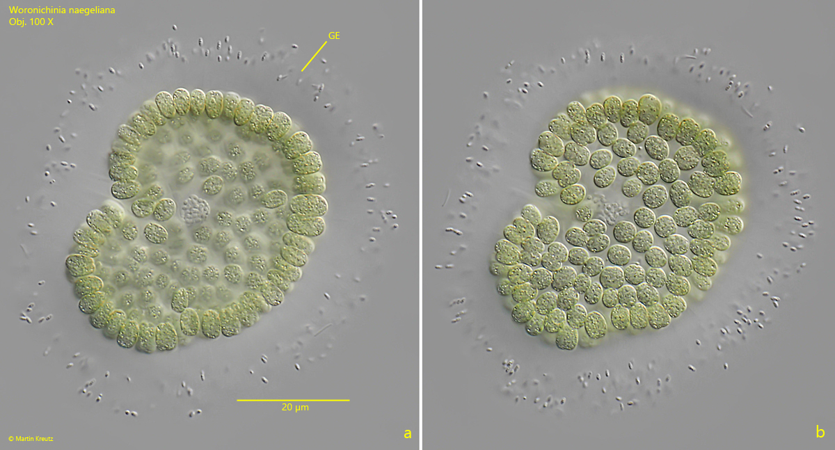
Fig. 2 a-b:Woronichinia naegeliana. D = 54 µm (of colony). The colony as shown in fig. 1 a-b in DIC. Note the distinct gelatinous layer of the colony. The stalks conecting the cells can only be seen after staining. Obj. 100 X.