two rings of fine, granulated pseudopodia around the base of the anterior flagellum and in the posterior third
posteriorly a thin stalk, trailing behind or attached to the substrate
one spherical nucleus in mid-body
2 contractile vacuoles near surface
Actinomonas mirabilis
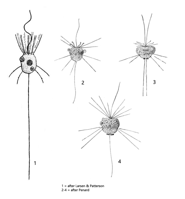
Actinomonas mirabilis is very similar to Pteridomonas pulex, but Actinomonas mirabilis has two rings of pseudopodia that arise more or less vertically from the cell surface. The anterior ring originates at the base of the flagellum, while the second ring is located in the posterior third of the body (s. figs, 1 a-c and 2). The pseudopodia are rigid and finely granulated (s. fig. 2). This flagellate was described as Pteridomonas scherffeli by Penard in 1921, but was considered synonymous with Actinomonas mirabilis by Larsen and Patterson in 1985.
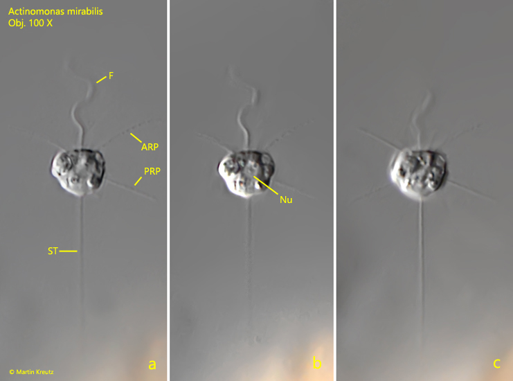
Fig. 1 a-c:Actinomonas mirabilis. L = 8.4 µm. Three focal planes of a specimen attached to a detritus flake. Note the two rings of pseudopodia arising at the anterior end (ARP) and in the posterior third of the cell (PRP). F = flagellum, ST = stalk. Obj. 100 X.
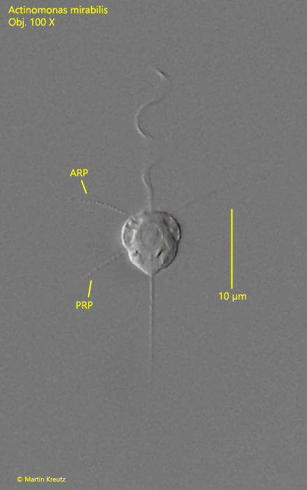
Fig. 2:Actinomonas mirabilis. L = 8.7 µm. A second specimen with the anterior ring of pseudopodia (ARP) and the posterior ring of pseudopodia (PRP). Obj. 100 X.