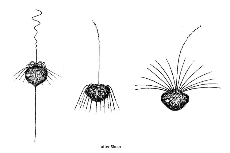
one ring of fine pseudopodia around the base of the anterior flagellum
posteriorly a thin stalk, trailing behind or attached to the substrate
one spherical nucleus in mid-body
2–3 contractile vacuoles near surface
Pteridomonas pulex
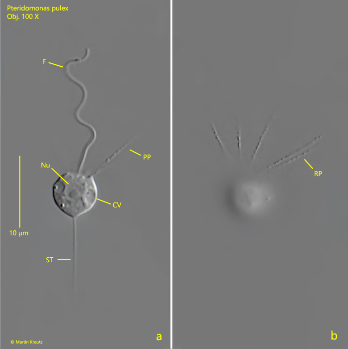
Pteridomonas pulex is a very small, colorless flagellate that is difficult to find in samples under the coverslip. It can be better observed on floating coverslips, on which Pteriodomonas pulex sometimes settles. The exact structure of the flagellate can only be seen at high magnification. At the base of the apical flagellum a ring of fine granulated pseudopodia arises (s. fig. 1b). This feature distinguishes Pteridomonas pulex from the similar species Actinomonas mirabilis (Kent, 1880), which has two such rings. A fine stalk of cytoplasm arises from the posterior end of Pteridomonas pulex (s. fig. 1a). With this stalk the flagellate can attach itself to the substrate (or coverslip). In freely swimming specimens this stalk is sometimes absent.
A virtually identical species was found in saltwater and described as Pteridomonas danica by Patterson and Fenchel (1985). I could not see any difference in the description of Pteridomonas danica and Pteridomonas pulex. Likely this flagellate occurs in both saltwater and freshwater that is Pteridomonas danica and Pteridomonas pulex are identical species.
Fig. 1 a-b:Pteridomonas pulex. L = 6.8 µm. Two focal planes of a specimen attached to a floating coverslip. Note the ring of pseudopodia (RP) arising at the base of the flagellum (F). CV = contractile vacuole, Nu = nucleus, PP = pseudopodium, ST = stalk. Obj. 100 X.