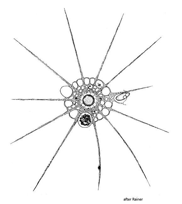
body spherical, diameter 20–90 µm (commonly 40–50 µm)
ectoplasm vacuolated
endoplasm granulated with numerous small vacuoles
a large central nucleus, diameter 10–15 µm
one contractile vacuole protrudes above surface before contracting
axopodia relatively strong, tapering to distal end
microtubules of the axopodia arsing from membrane of the nucleus
formation of colonies and feeding communities
Actinophrys sol
Actinophrys sol belongs to the most common heliozoans. The species can also occur in masses. The species can be easily recognized by the strongly vacuolated cytoplasm and the pointed axolpodia. Compared to Actinosphaerium eichhornii, Actinophrys sol is much smaller and, most importantly, has only one centrally located nucleus. The internal structure of Actinosphaerium sol is best examined on starving specimens where no phagocytosed food interferes (s. figs. 2, 3 and 4). In such specimens, one can see very nicely how the microtubules, which form the axial rod of the axopodia, extend from the surface of the nucleus (s. figs. 3 and 4). Actinophrys sol can capture very large prey organisms and also forms feeding communities.
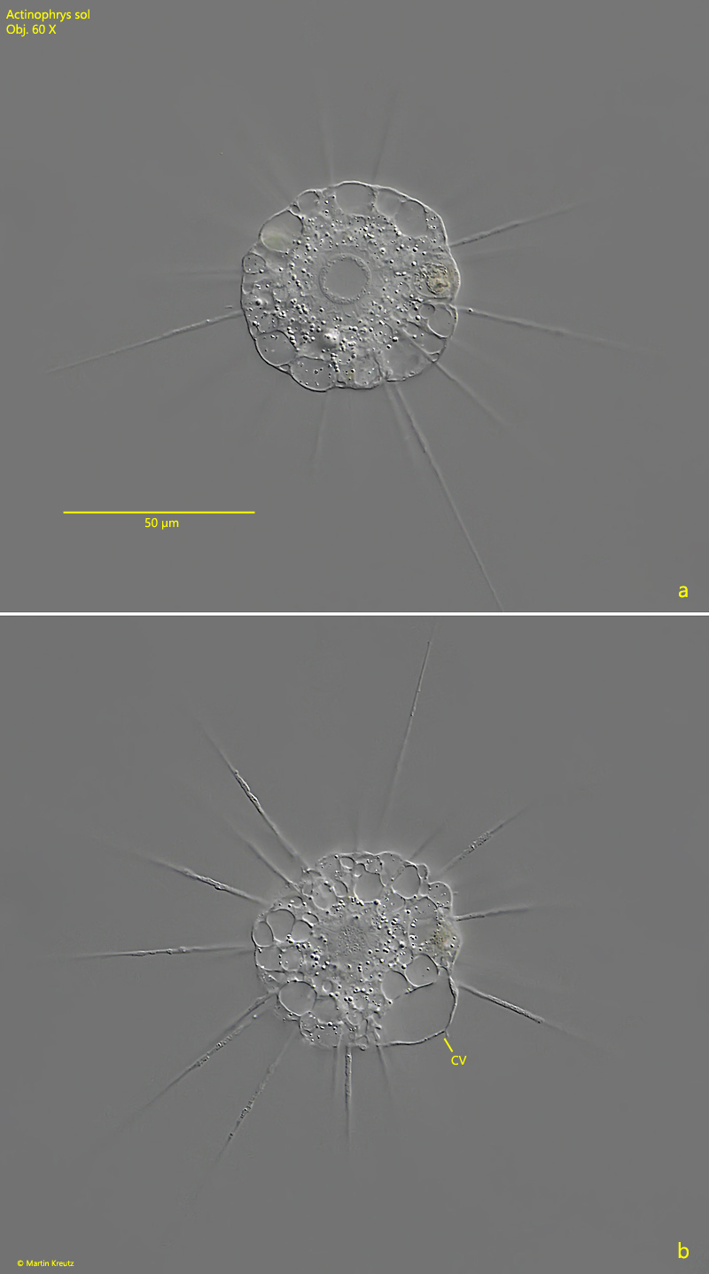
Fig. 1 a-b:Actinophrys sol. D = 52 µm. Two focal planes of an extended specimen. Note the contractile vacuole (CV) protruding above the cell surface. Obj. 60 X.
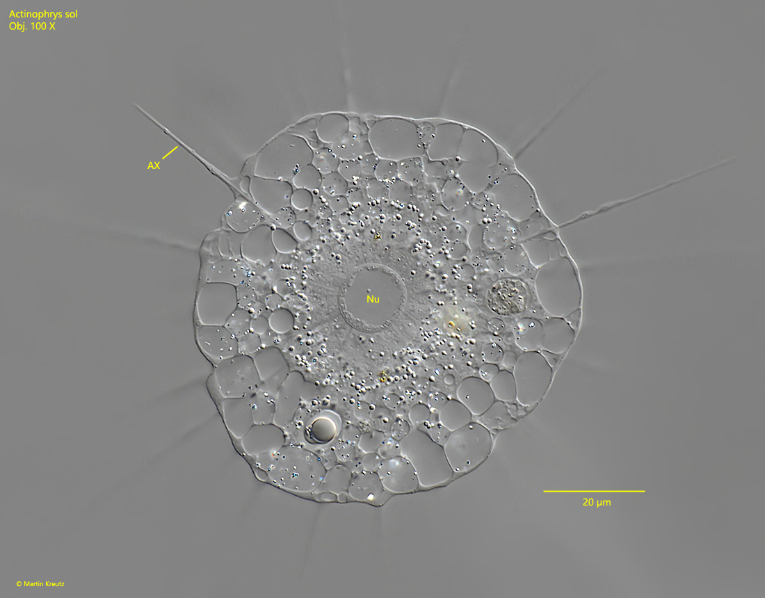
Fig. 2:Actinophrys sol. A squashed specimen with the central nucleus (Nu). AX = Axopodium. Obj. 100 X.
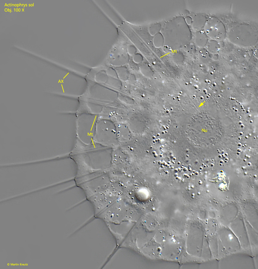
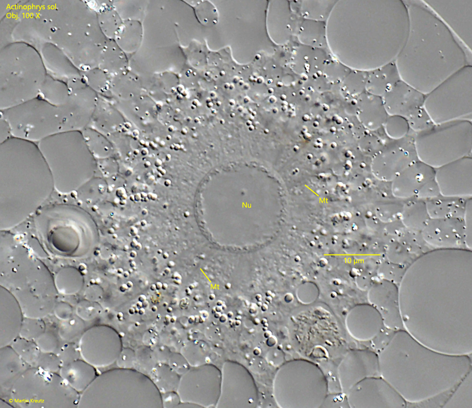
Fig. 3:Actinophrys sol. The microtubules (Mt) of the axopodia (AX) arising from the surface of the nucleus (Nu, arrow). Obj. 100 X.
Fig. 4:Actinophrys sol. A different focal plane of the specimen shown in fig. 3. The microtubules (Mt) arising from the surface of the nucleus (Nu). Obj. 100 X.