So far I have only found Aphanocapsa parasitica in the plankton in the pond of the convent Hegene. Here I found this interesting cyanobacteria in the loricae of Dinobryon stipitatum. I have never been able to find this species in my other sampling sites.
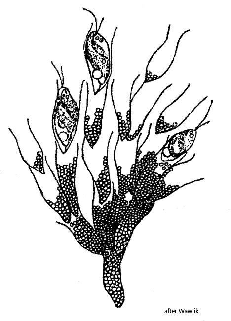
Aphanocapsa parasitica occurs both epiphytically on algae and in the posterior part of the loricae of Dinobryon. It is difficult to identify the epiphytic form, as the species of Aphanocapsa are mainly defined by the size of the spherical cells. However, if Aphanocapsa parasitica is present in the loricae of Dinobryon, the identification is unambiguous. Whether it is really a parasitic relationship can be doubted, because “infested” colonies of Dinobryon do not seem to be affected in any way. The loricae of Dinobryon are probably colonized in order to remain in the upper, light-flooded water layer with the mobile colonies of Dinobryon. Metabolic products of the flagellate may also be used.
In my population, the cells of Aphanocapsa parasitica had a diameter of 1.3–1.5 µm. The cytoplasm appeared homogeneous. The blue-greenish color was very pale. I could not detect any mucus formation. In all cases of Dinobryon that were colonized with Aphanocapsa parasitica, there was a kind of sediment in the pointed ends of the loricae. Sometimes this “sediment” was hyaline, sometimes structured. I could not observe how the cells of Aphanocapsa parasitica can pass the flagellate to get in the posterior part of the lorica. However, the process seems to be very effective because almost all Dinobryon stipitatumloricae were colonized with Aphanocapsa parasitica.
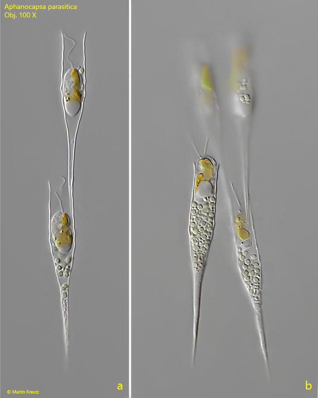
Fig. 1 a-b:Aphanocapsa parasitica. D = 1.3–1.5 µm. Some colonies of the spherical cells in the posterior parts of the loricae of Dinobryon stipitatum. Obj. 100 X.
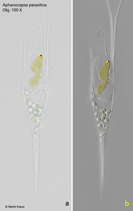
Fig. 2 a-b:Aphanocapsa parasitica. D = 1.3–1.5 µm. A colony of only few cells in the posterior part of the lorica of Dinobryon stipitatum in brightfield illumination (a) and DIC (b). Note the pale blue-green color of the cells. Obj. 100 X.
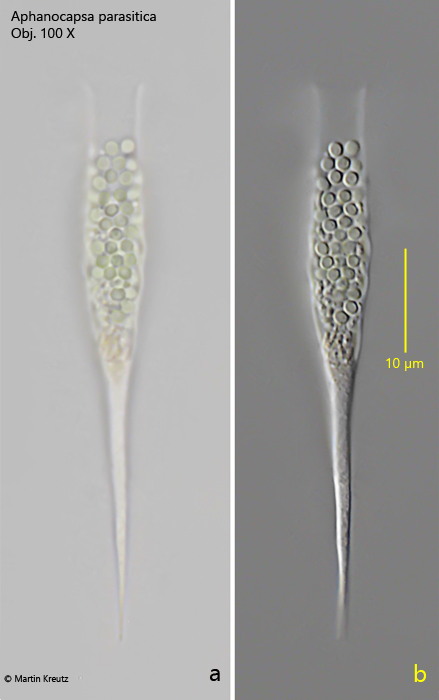
Fig. 3:Aphanocapsa parasitica. D = 1.3–1.5 µm. An empty lorica of Dinopryon stipitatum in brightfield illumination (a) and DIC (b) with a colony of about 50 cells. Obj. 100 X.