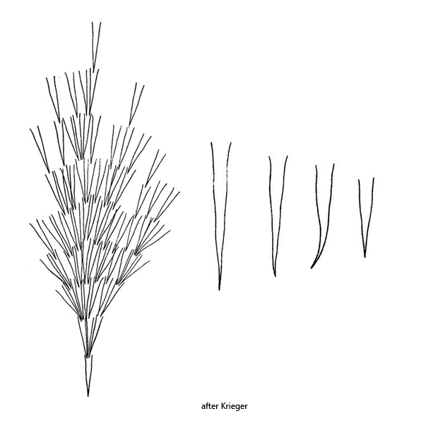
lorica vase-shaped, long and straight with long tapering portion
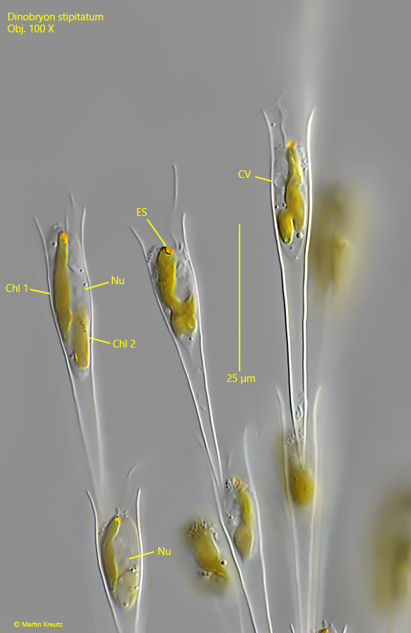
cells attached with a tapered stalk of cytoplasm to the posterior part of the lorica
two golden brown colored chloroplasts
the anterior chloroplast with an eyespot
two flagella of different lengths
one spherical nucleus between chloroplasts
two contractile vacuoles in midbody
length of lorica 35–70 µm
cells forming a branched colony, branches long and straight
colony up to 500 µm long
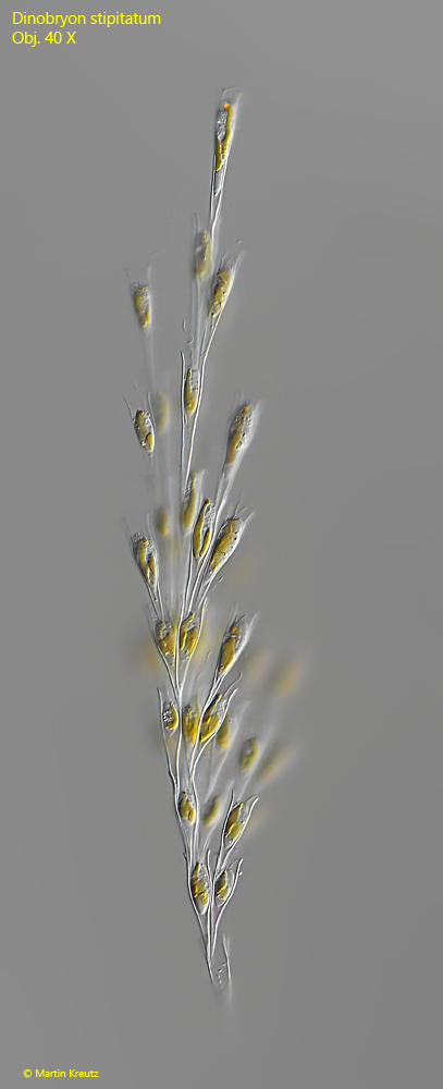
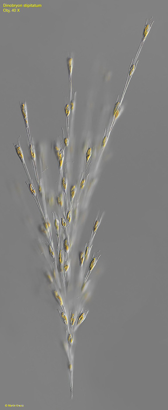
Dinobryon stipitatum
I have found Dinobryon stipitatum only very rarely and exclusively in the plankton of Lake Constance. The lorica of this species is very slender with a long, tapered end. The anterior third is only slightly bulbous or parallel sided and the opening of the lorica is somewhat widened. Due to the slender shape of the loricae, the branches of the colonies also appear slender and straight. The colonies can grow up to 500 µm long. The cells are typical of the genus Dinobryon and do not differ from Dinobryon sertularia or Dinobryon divergens.
Fig. 1:Dinobryon stipitatum. L = 420 µm (of colony). A freely floating colony. Obj. 40 X.
Fig. 2:Dinobryon stipitatum. L = 490 µm (of colony). A second freely floating colony. Obj. 40 X.
Fig. 3:Dinobryon stipitatum. L = 47 µm (of lorica). The loricae are straight and slender with a tapered end. The distal end is slightly widended. The cells are about 20 µm long. CHl 1-2 = chloroplasts, CV = contractile vacuole, ES = eyespot, Nu = nucleus. Obj. 100 X.