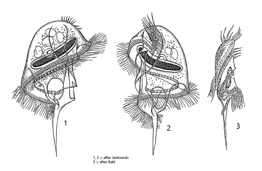
body medusoid, slightly flattened, with a prominent dome and a long caudal spine
length 150–200 µm
on dorsal side of the dome two rows of cirri
without somatic cilia except for a tuft of cilia at the base of the caudal spine
perizonal stripe consists of 5 ciliary rows
adoral zone runs in a furrow and encircles the body spirally
mouth opening in mid-body, cytopharynx directed anteriorly
one elongated or vermiform macronucleus with one adjacent, spherical micronucleus
one contractile vacuole located at the base of caudal spine
Caenomorpha sapropelica
Caenomorpha sapropelica is very common in most of my sampling localities. Especially in the Purren pondCaenomorpha sapropelica can be found reliably. The anterior part of the body, with apical dome and the circumferential adoral zone occupies about 2/3 of the body length, while the caudal spine occupies about 1/3 of the body length. Caenomorpha sapropelica is a fast swimmer, rotating around its longitudinal axis. The very long cilia, which originate dorsally on the apical dome, perform a rudder-like movement in the process. I could never observe a thigmotactic attachment of these long cilia to the subtrate. They are also not fused to form cirri as in Caenomorpha medusula, but rather soft and flexible. The mouth opening is located approximately at the level of the contractile vacuole. However, the cytopharynx is directed towards the anterior end (s. fig 5 b). The adoral zone begins dorsally, to the left of the two rows of apical rows of cilia, and completely encircles the body to the mouth opening. Here, the adoral membranelles lie in a furrow covered by the overhanging margin of the apical dome (s. fig. 5b). The perizonal stripe runs in parallel to the adoral zone on the distal edge of the apical dome (s. fig. 4a and 5a). At the base of the terminal spine there is a tuft of cilia (s. fig. 2 b and 3 b). In the food vacuoles I could find mainly colorless bacteria and yellowish sulfur bacteria. In the apical dome there is an accumulation of highly refractive granules, but these are only visible in the squashed specimen. In the cytoplasm there are also highly refractive spheres (oil?) and numerous symbiotic bacteria (s. fig. 6).
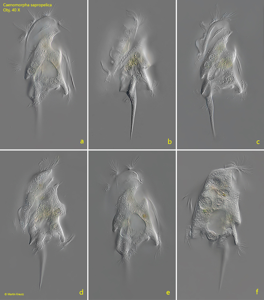
Fig. 1 a-f:Caenomorpha sapropelica. L = 157 µm. A freely swimming specimen from ventral (a, c, e) and from right (b, d, f). Obj. 40 X.
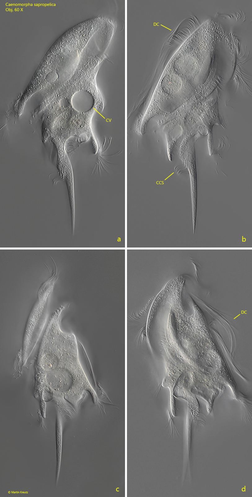
Fig. 2 a-d:Caenomorpha sapropelica. L = 160 µm. A second freely swimming specimen from right (a, b, c) and left (d). Note the long cilia arising at the dorsal side of the apical dome (DC). CV = contractile vacuole, CCS = tuft of cilia at the base of the caudal spine. Obj. 60 X.
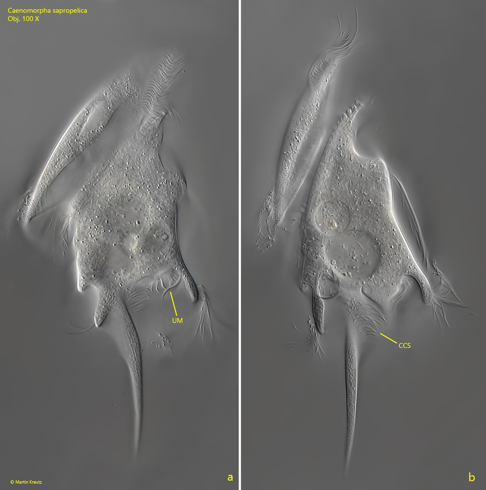
Fig. 3 a-b:Caenomorpha sapropelica. L = 164 µm. A third freely swimming specimen from right. CSS = tuft of cilia at the base of the caudal spine, UM = undulating membrane. Obj. 100 X.
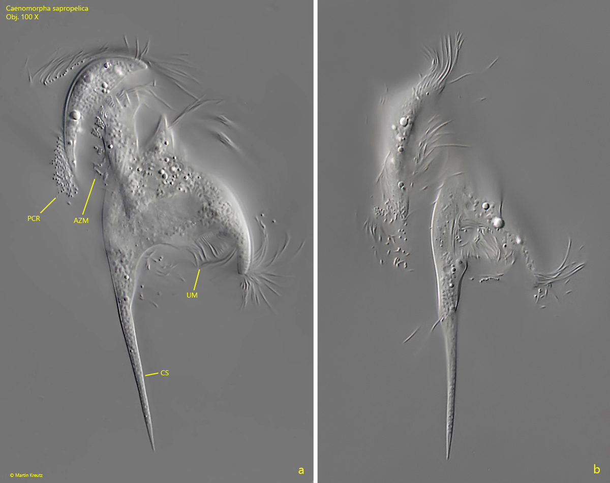
Fig. 4 a-b:Caenomorpha sapropelica. Two optical sections of a slightly squashed specimen from right. AZM = adoral zone of membranelles, CS = caudal spine, PCR = perizonal cilia row, UM = undulating membrane. Obj. 100 X.
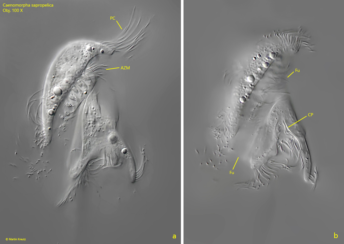
Fig. 5 a-b:Caenomorpha sapropelica. Two optical sections of a slightly squashed specimen from right. AZM = adoral zone of membranelles, CP = cytopharynx, CS = caudal spine, Fu = furrow beneath the apical dome, PC = perizonal cilia. Obj. 100 X.
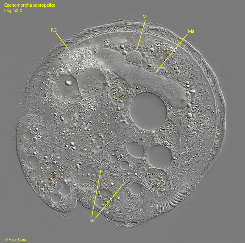
Fig. 6:Caenomorpha sapropelica. The vermiform macronucleus (Ma) and the adjacent micronucleus (Mi) in a strongly squashed specimen. SB = symbiotic bacteria in the cytoplasm, RG = aggregation of refractive granules located in the apical dome. Obj. 100 X.