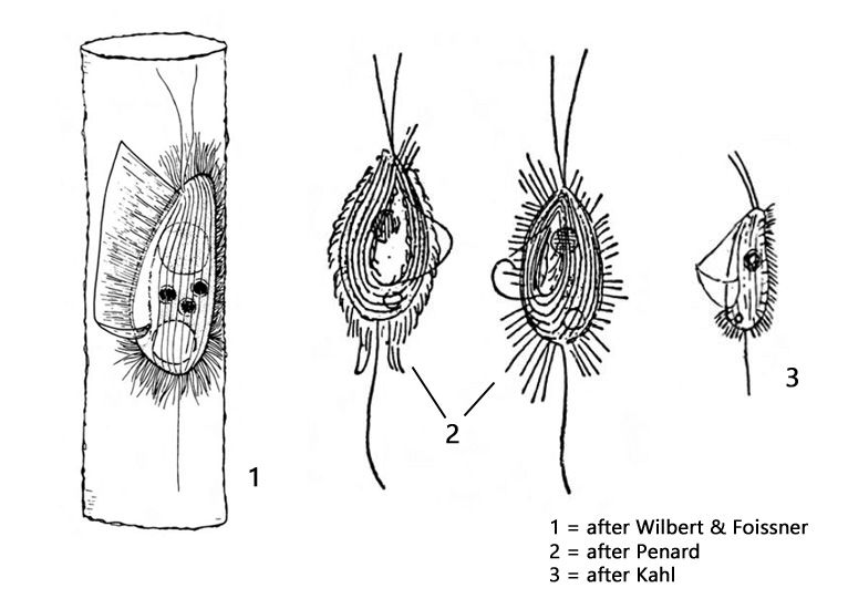
lorica may have paralles sides or a central bulbous region
prominent L-shaped undulating membrane
macronucleus in anterior half
extrusomes present
two elongated apical cilia (hard to see)
long caudal cilium
CV terminal
Calyptotricha lanuginosa
Calyptotricha lanuginosa builds a lorica similar to that in Calyptotricha pleuronemoides, but can be easily distinguished from it by the absence of symbiotic algae. In addition, Calyptotricha lanuginosa is slightly smaller and the caudal cilium is usually slightly longer. Apart from this, the characteristics are largely to the situation in Calyptotricha pleuronemoides. I found Calyptotricha lanuginosa so far exclusively in Simmelried.
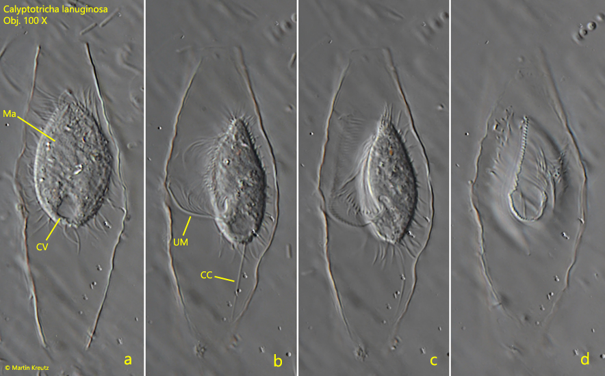
Fig. 1 a-d:Calyptotricha lanuginosa. L = 27 µm. A freely rotating specimen in the lorica. a) dorsal view; b, c) lateral view from left; d) ventral view. CV = contractile vacuole, Ma = macronucleus, UM = undulating membrane. Obj. 100 X.
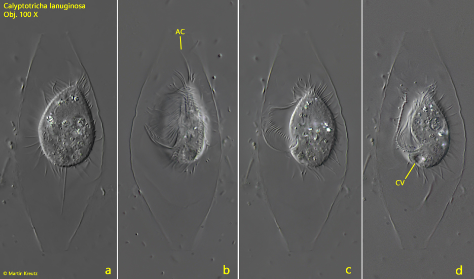
Fig. 2 a-d:Calyptotricha lanuginosa. L = 24 µm. A second freely rotating specimen in the lorica. a) dorsal view; b, c = lateral view from left; d) ventral view. AC = apical cilia, CV = contractile vacuole. Obj. 100 X.
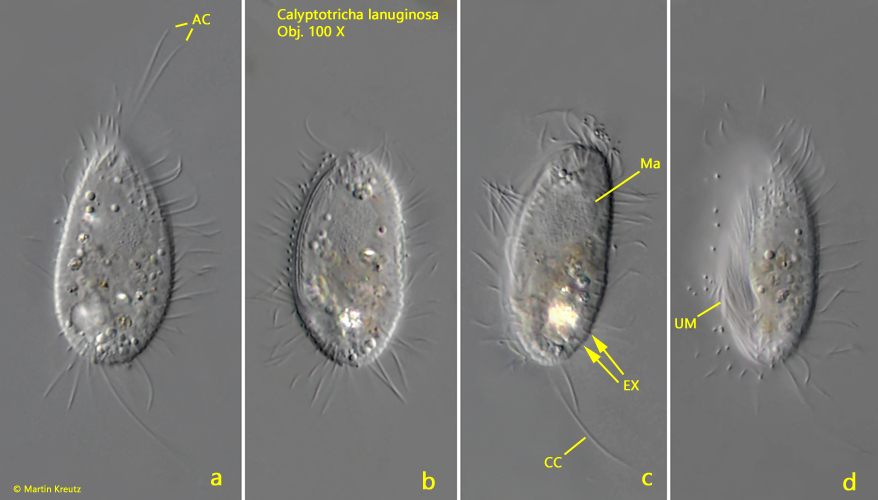
When disturbed (e.g. coverslip pressure), Calyptotricha lanuginosa often leaves the lorica and swims around. When such specimens are found, they are easily confused with other pleurostomatid ciliates such as Cyclidium. However, they can be clearly identified by the typical 2 elongated apical cilia.
Fig. 3 a-d:Calyptotricha lanuginosa. L = 30 µm. A freely swimming specimen outside of the lorica. AC = apical cilia, CC = caudal cilium, EX = extrusomes, Ma = macronucleus. Obj. 100 X.