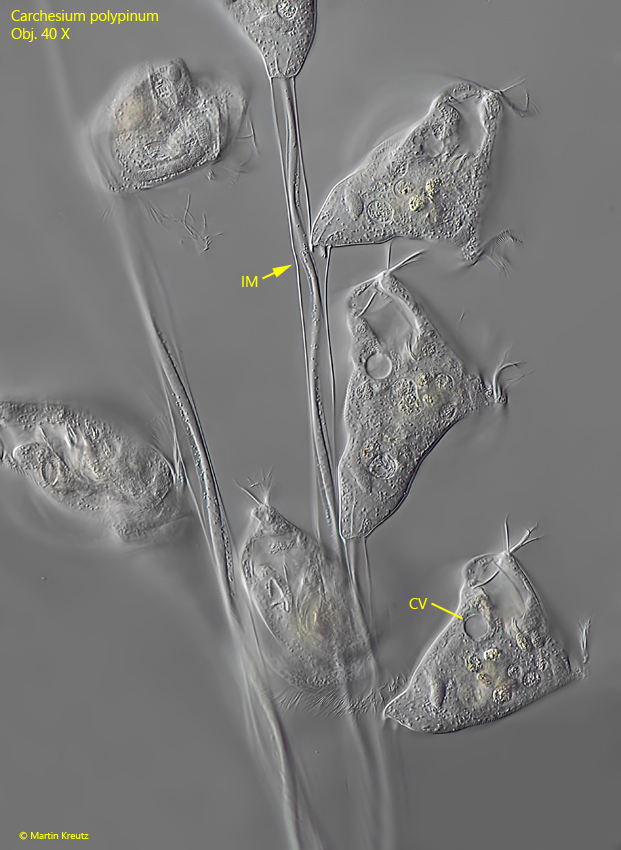
zooids elongated bell-shaped, often tilted to the side
length of zooids 80–140 µm
zooids contract almost spherically
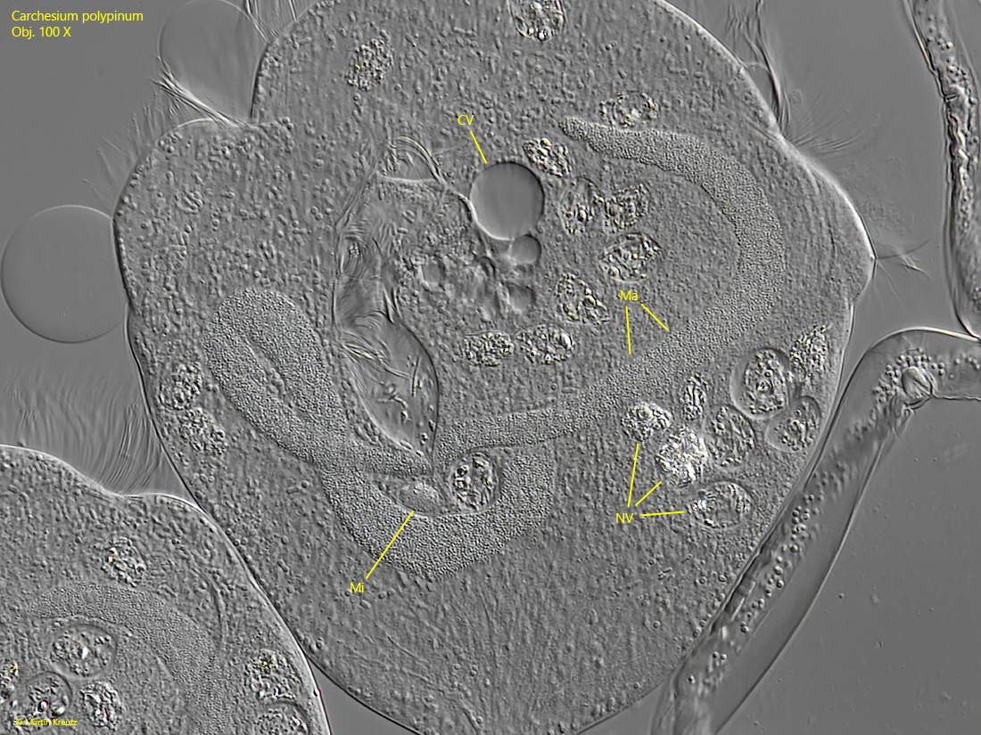
macronucleus J-shaped in the longitudinal axis
one micronucleus
one contractile vacuole
peristome collar 60–135 µm in diameter
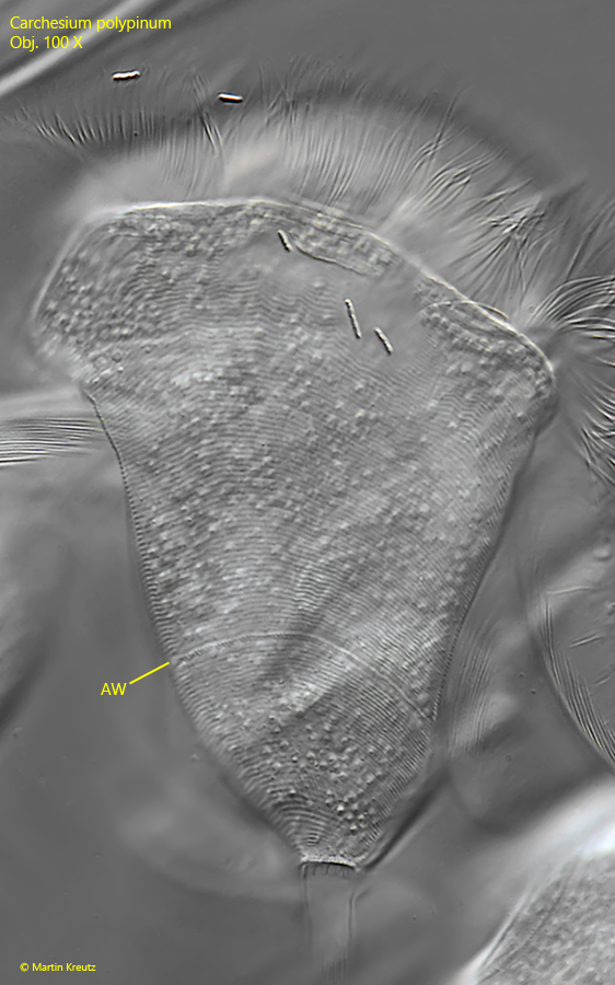
pellicle finely striated
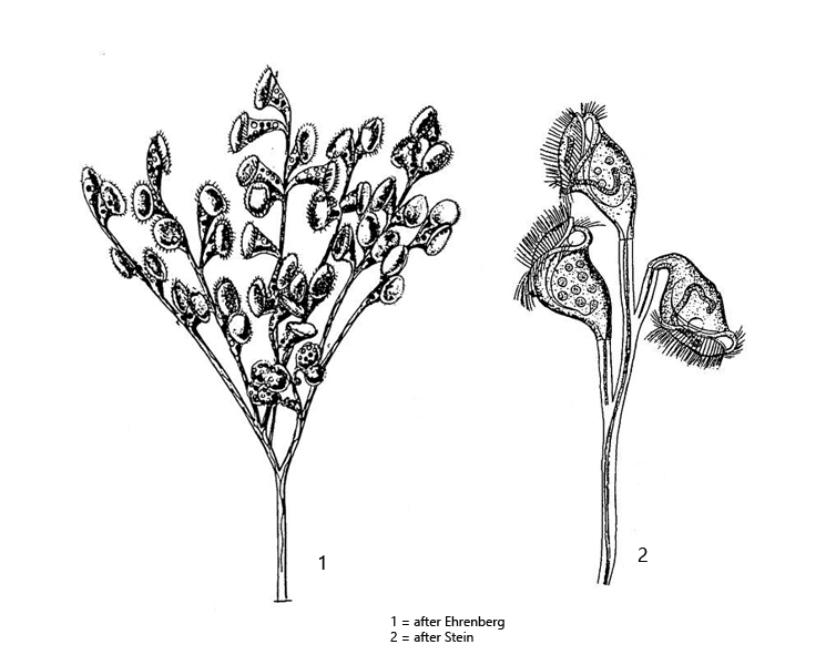
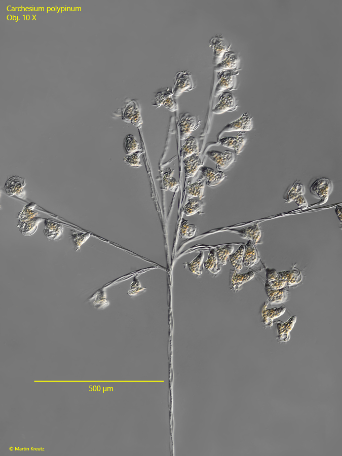
colonies tree-shaped with branched stalks, up to 2 mm heigh
lower part of the colonies is an unbranched main stem
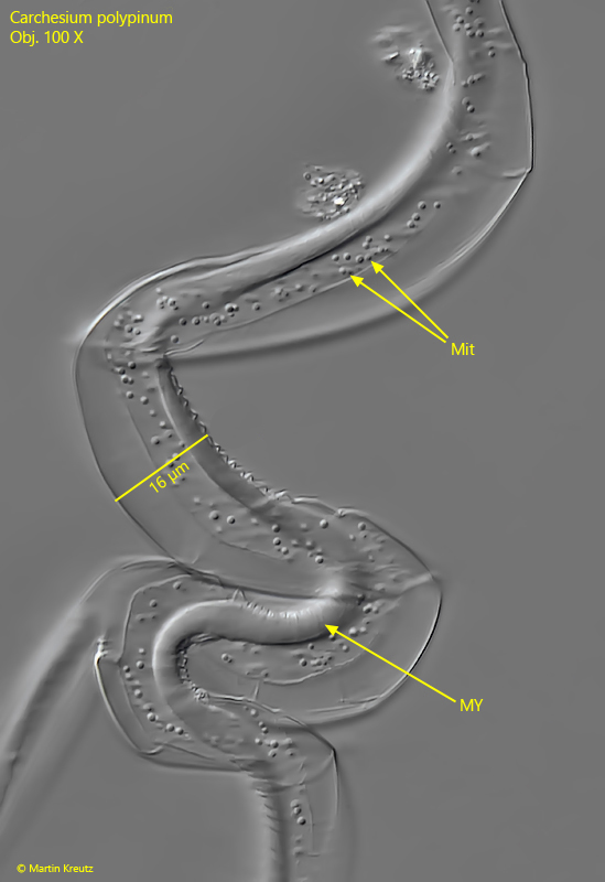
stalks contract in tight helix
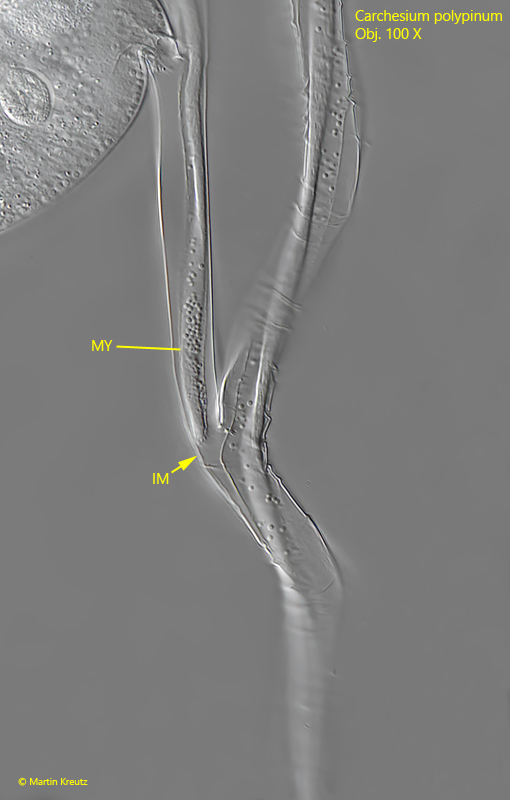
myonemes in the branches are interrupted
Carchesium polypinum
I find Carchesium polypinum mainly in old samples from the Simmelried. The colonies of this pertrich ciliate are easy to recognize by the tree-like structure and the non-branched stalk. Carchesium polypinum can be easily distinguished from other colony-forming peritrich ciliates, whose peduncles also contract helically, by the interrupted myonemes at the branching points (s. figs. 3, 4 and 5). This interruption of myonemes causes zooids and also individual branches of colonies to contract independently. In the similar genus Zoothamnium, the myonemes are not interrupted, causing the entire colony to always contract synchronously. A distinguishing feature from Vorticella species is the stalk thickness. In Vorticella, the stalk is always < 10 µm thick, whereas in Carchesium polypinum it is thicker than 10 µm (s. fig. 6). Comparable Epistylis species (e.g. Epistylis procumbens) do not have a central myoneme in the stalk and therefore cannot contract.
Fig. 1: Carchesiumpolypinum. Overview of a colony with a length of 1600 µm. Obj. 10 X.
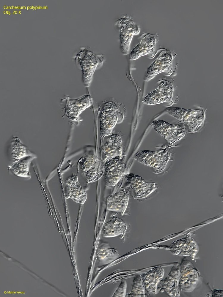
Fig. 2: Carchesiumpolypinum. Detail of the colony shown in fig. 1. Obj. 20 X.
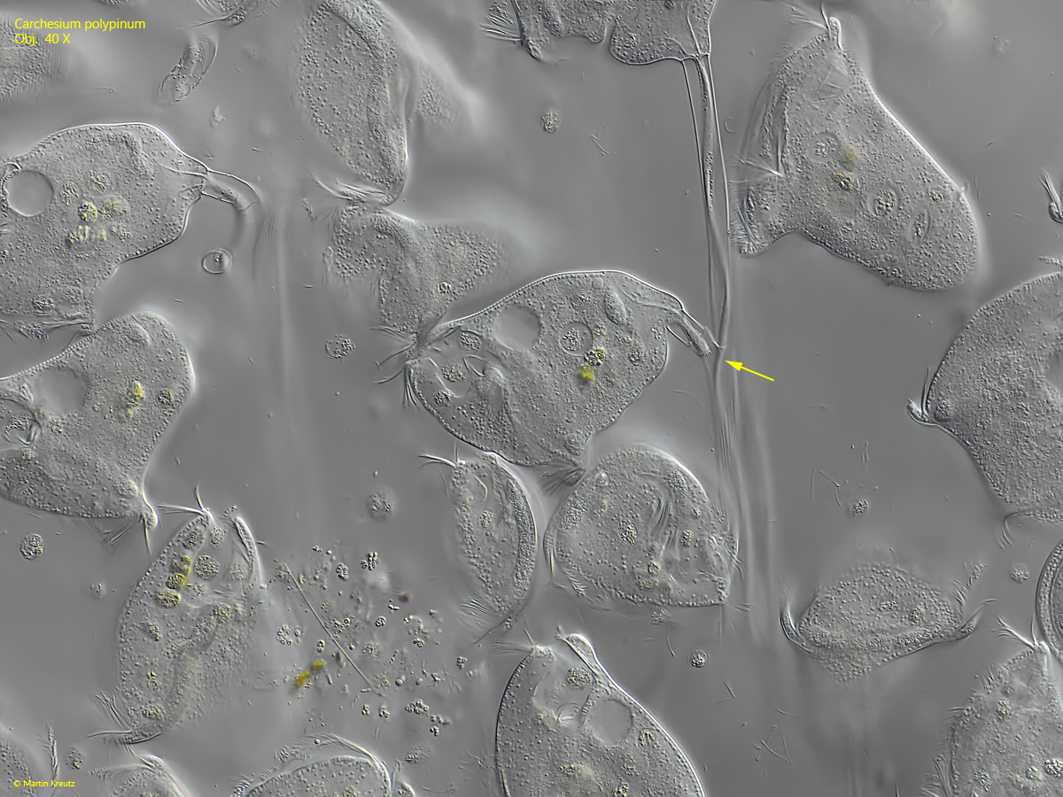
Fig. 3: Carchesiumpolypinum. L = 90–106 µm (of zooids). The zooids of the colony shown in fig. 1. Note the interrupted myoneme (IM) at the branch of the stalk. Obj. 40 X.
Fig. 4: Carchesiumpolypinum. The interrupted myoneme (arrow) at the branch of the stalk in a slightly squashed colony. Obj. 40 X.
Fig. 5: Carchesiumpolypinum. The interruption of the myoneme (IM) in detail. My = myoneme of the stalk. Obj. 40 X.
Fig. 6: Carchesiumpolypinum. The spiralized stalk with the central myoneme (MY). The myoneme is surrounded by numerous mitochondria (Mit). Obj. 100 X.
Fig. 7: Carchesiumpolypinum. The fine striation of the pellicle. In this specimen, there are 15 lines per 10 µm. AW = aboral ciliary wreath. Obj. 100 X.
Fig. 8: Carchesiumpolypinum. The J-shaped macronucleus (Ma) and the micronucleus (Mi) in a strongly squashed specimen. CV = contractile vacuole. Obj. 100 X.