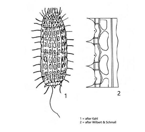
apical mouth opening with basket of pharyngeal trichites
posteriorly mostly 3 strong spines
armour composed of 6 rings, each with 12–14 plates
plates with a variable number of half and whole “windows”
“windows” kidney-shaped
macronucleus spherical in mid-body with one adjacent micronucleus
contractile vacuole sub-terminal
one caudal cilium
no symbiotic algae
Nolandia nolandi
Nolandia nolandi was first described by Kahl as Coleps nolandi. Based on morphological characteristics and genetic studies, the species was transferred to the genus Nolandia by Small & Lynn in 1985.
At low magnifications Nolandia nolandi is difficult to distinguish fromColeps hirtus var. minor, because both species have a similar size and body shape. Only at high magnification can the shape of the “windows” in the armour be seen, which is essential for identification. In Nolandia nolandi the windows are narrow and kidney shaped. In my population I could detect 2 whole “windows” and 2 half “windows” per half cell. However, the number of half and whole “windows” per half cell varies and is therefore not a definite identification characteristic. The decisive factor is the shape of the “windows”.
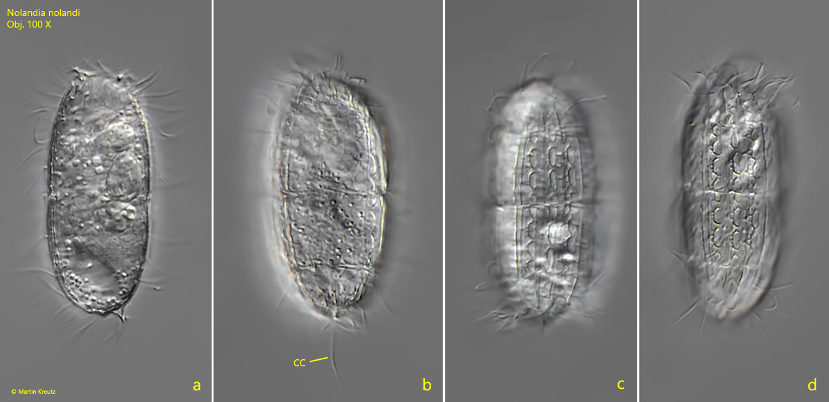
Fig. 1 a-d:Nolandia nolandi. L = 50 µm. Different focal planes of a freely swimming specimen. CC = caudal cilium. Obj. 100 X.
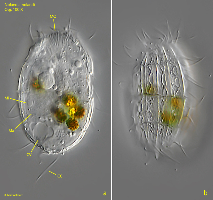
Fig. 2 a-b:Nolandia nolandi. L = 36 µm. Two focal planes of a slightly squashed specimen. CC = caudal cilium, CV = contractile vacuole, Ma = macronucleus, Mi = micronucleus, MO = mouth opening. Obj. 100 X.
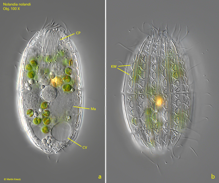
Fig. 3 a-b:Nolandia nolandi. L = 54 µm. Two focal planes of a second squashed specimen. Note the kidney-shaped “windows” (KW). CV = contractile vacuole, CP = cytopharynx, Ma = macronucleus. Obj. 100 X.