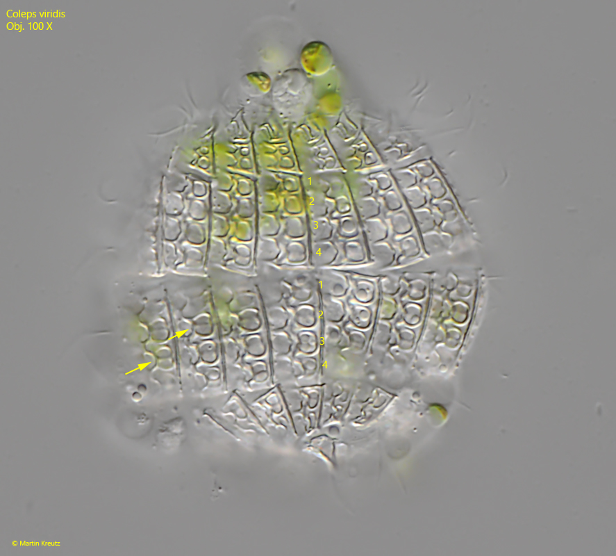
anterior and posterior main plate with 4 “windows” each
shape of the “windows” in the armour pretzel-shaped
contractile vacuole terminal
macronucleus spherical
one caudal cilium
1-4 short spines at posterior end
No drawings from previous authors available.
The species Coleps viridis was established by Ehrenberg in 1831, but later considered as a form of Coleps hirtus by Kahl and named Coleps hirtus viridis. It was not until 2021 that Pröschold et al. subjected Coleps hirtus viridis to morphological and genetic analyses, including symbiotic algae. As a result of the study, Pröschold et al. re-established the original name Coleps virdis and were able to identify the symbiotic algae as Micractinium conductrix.
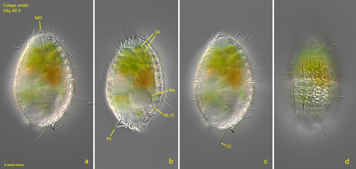
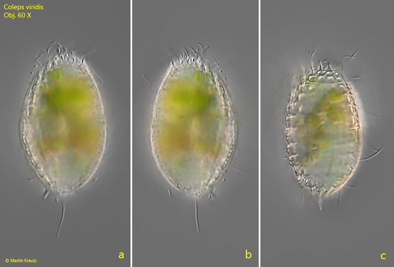
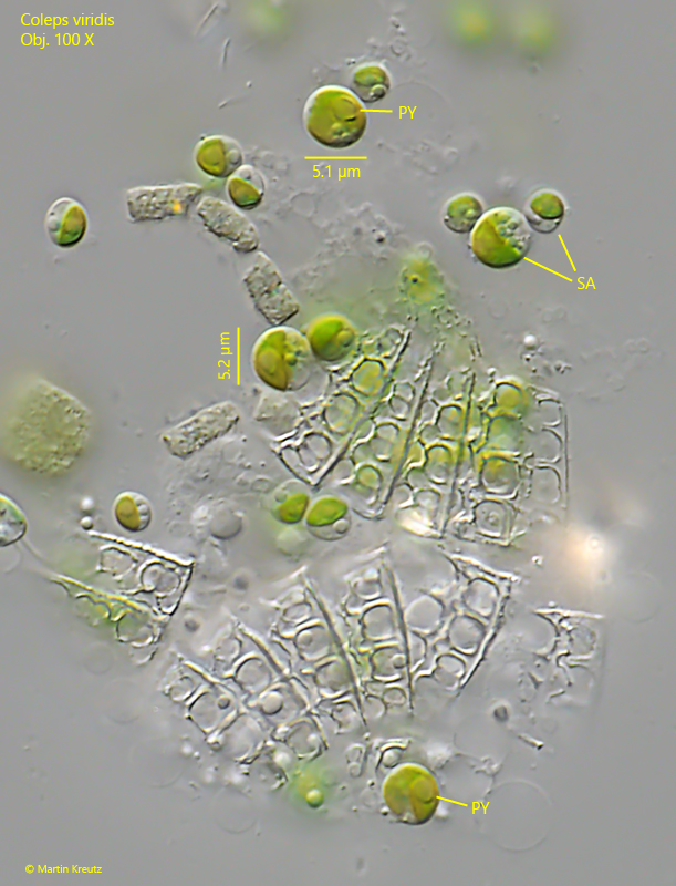
In my population all specimens of Coleps viridis were smaller than 50 µm. Because of the small size the species is probably often overlooked or confused with Coleps hirtus or Coleps hirtus var. minor. At low magnifications the cells appear opaque black. Only at higher magnifications does the green coloration become visible due to the symbiotic algae (s. figs. 1 a-d and 2 a-c). I could count 15–30 symbiotic algae in each cell. The algae cells have a cup-shaped chloroplast with a pyrenoid, are spherical and have a diameter of about 5.0 -5.4 µm (s. fig. 3). Thus, they correspond to the description of Micractinium conductrix by Pröschold et al. Coleps virdis has one caudal cilium (s. figs. 1 c and 2 a-b) and the armour is of the Coleps hirtustype. That means each main plate has 4 “windows” which are pretzel-shaped (s. fig. 4).
Fig. 1 a-d:Coleps viridis. L = 47 µm. A freely swimming specimen. CC = caudal cilium, Ma = macronucleus, Mi = probably the micronucleus, MO = mouth opening, PS = posterior spine, SA = symbiotic algae. Obj. 60 X.
Fig. 2 a-c:Coleps viridis. L = 44 µm. A second, freely swimming specimen. Obj. 60 X.
Fig. 3:Coleps viridis. A squashed specimen with the released symbiotic algae. The algae are spherical with a diameter of 5.0–5.4 µm, have a cup shaped chloroplast and one pyrenoid (PY). Obj. 100 X.
Fig. 4:Coleps viridis. In the anterior main plate as well as in the posterior main plate there are 4 “windows” (1–4) each. The “windows” are pretzel-shaped (arrows). Obj. 100 X.