I found my population of Colpoda cucullus in moss samples that came mainly from trees and walls. If the moos is soaked with rainwater in Petri dishes, a large population forms after a few days. Colpoda cucullus is actually the typical ciliate in hay infusions, where it practically always occurs. The habitats and reproduction strategies of Colpoda cucullus were studied in detail by Mueller & Muller (1970). According to this study, cysts of Colpoda cucullus are found on practically all green plants. They are activated by dew or rainwater. The life cycle of Colpoda cucullus is short enough for a complete reproduction cycle before the leaf surfaces become dry again. The cysts are transferred to other plants by pollen-collecting insects.
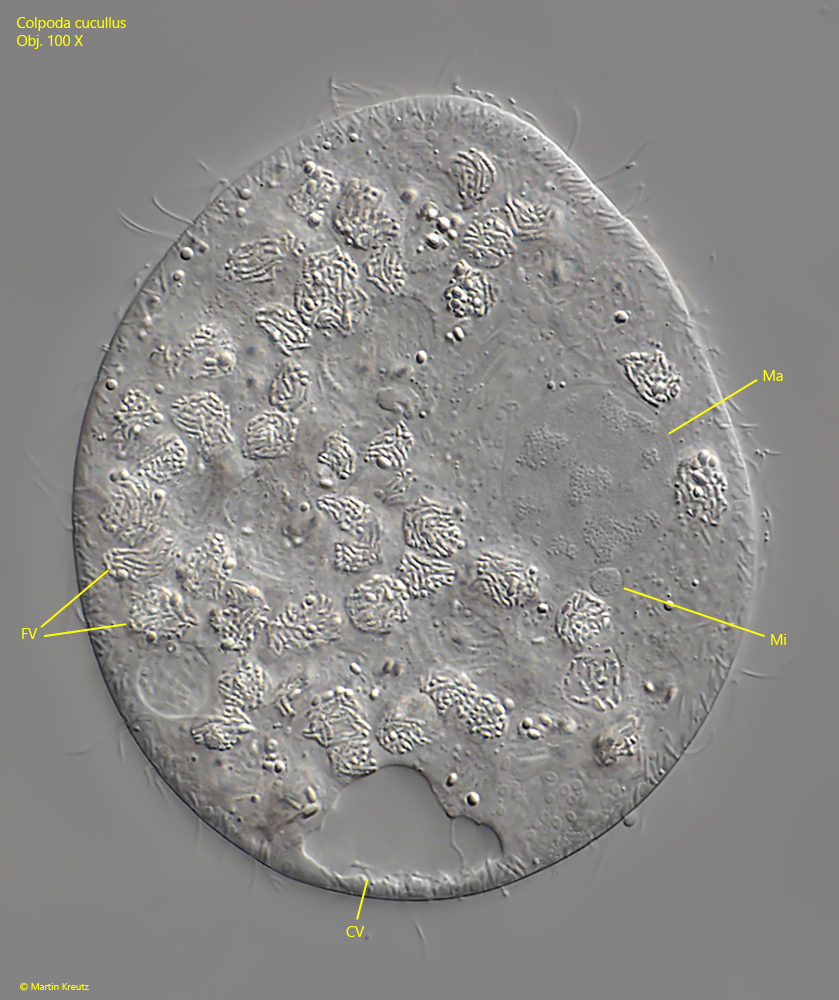
Colpoda cucullus is easily confused with Colpoda lucida in terms of size and shape. But Colpoda lucida has a clear, lightly-colored fringe of densely packed extrusomes, which makes it easy to distinguish. In my populations, the specimens of Colpoda cucullus were about 50-70 µm long, which is exactly in the lower range of the typical range given by Foissner. The macronucleus has no nucleoli, but is criss-crossed by chromatin islands, which can be clearly seen in squashed specimens (s. fig. 4). To the right and left of the mouth opening are two dense fields of cilia called polykinetids. In ventral view, these two polykinetids are clearly visible (s. fig. 3 a-b).
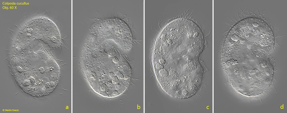
Fig. 1 a-d:Colpoda cucullus. L = 62 µm. Different focal planes of a freely swimming specimen from right. Obj. 60 X.
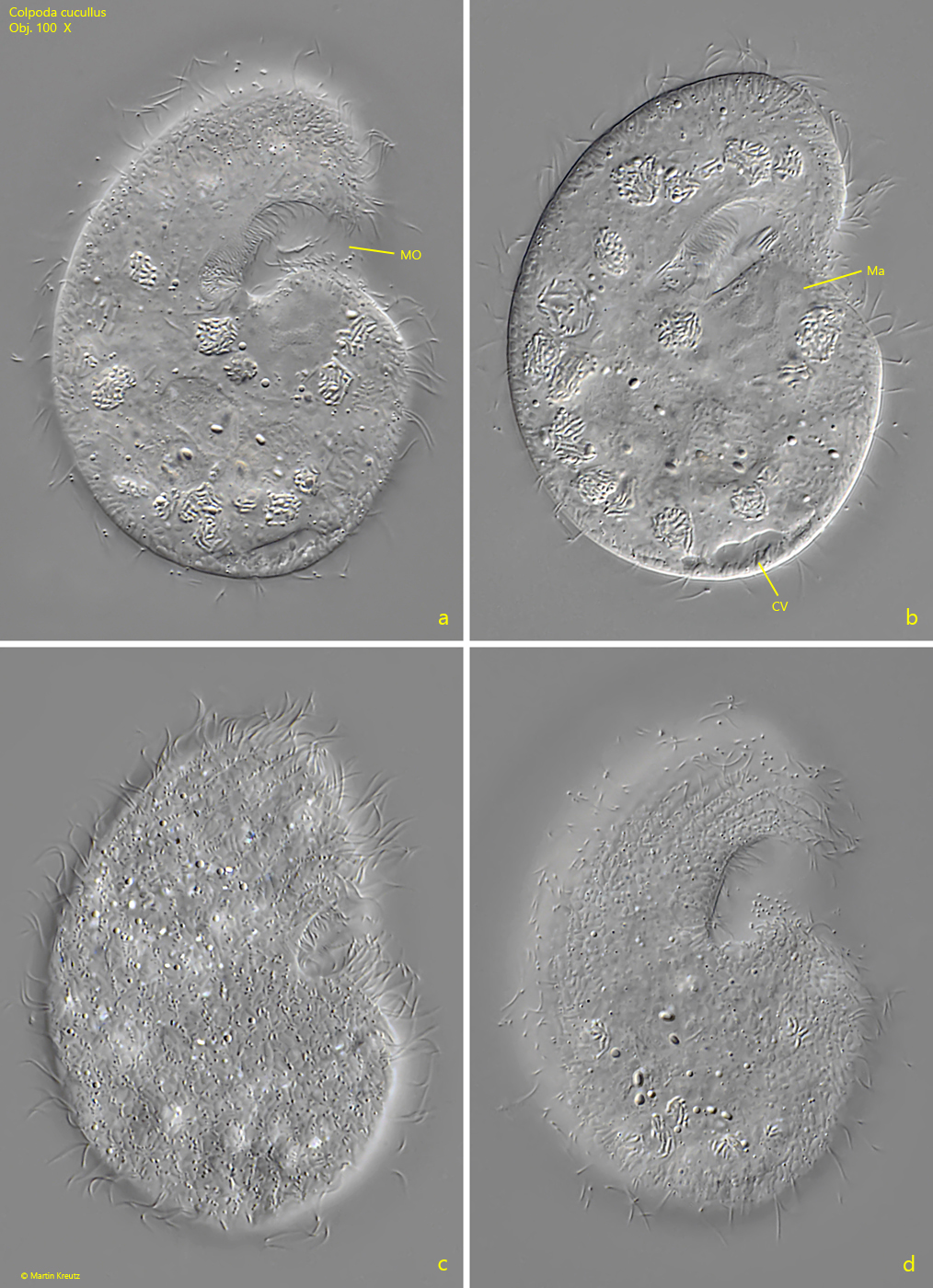
Fig. 2 a-d:Colpoda cucullus. L = 70 µm. Different focal planes of a slightly squashed specimen from right. Note the cloudy chromatin in the macronucleus (Ma). CV = contractile vacuole, Mo = mouth opening. Obj. 100 X.
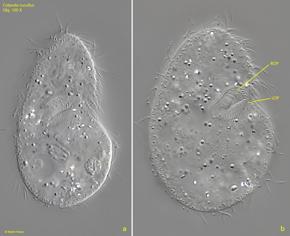
Fig. 3 a-b:Colpoda cucullus. L = 51 µm. A squashed specimen from ventral. Note the right oral polykinetid (ROP) and the left oral polykinetid (LOP). Obj. 100 X
Fig. 4:Colpoda cucullus. A strongly squashed specimen. CV = contractile vacuole, FV = food vacuoles, Ma = macronucleus with chromatin, Mi = micronucleus. Obj. 100 X
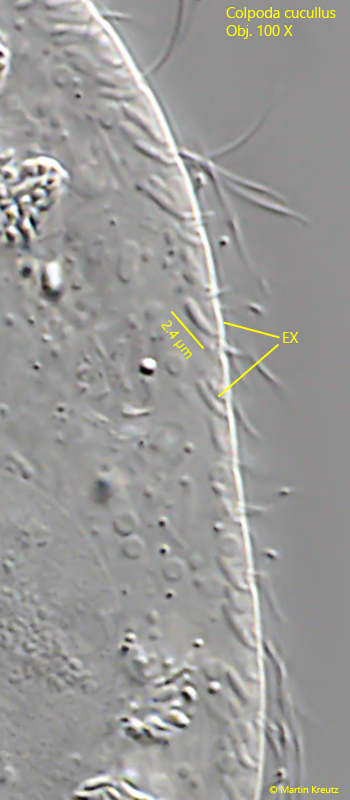
Fig. 5:Colpoda cucullus. The comma-shaped extrusomes (EX) in a strongly squashed specimen. Obj. 100 X