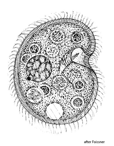
body broadly reniform, sometimes almost circular, laterally slightly flattened
length 70–110 µm, width 65–90 µm
semicircularly indented at the mouth opening
distinct fringe of extrusomes forms a cortical layer
extrusomes cylindrical to fusiform, 3–5 µm long
macronucleus globular to slightly ellipsoid with reticulate nucleolus
one micronucleus adjacent to macronucleus, about 6 x 4 µm
mouth opening with a right and left field of polykineties
ciliary rows consisting of paired cilia
contractile vacuole subterminal
no caudal cilia
Colpoda lucida
So far I have found Colpoda lucida exclusively in moss samples that I watered with rainwater.This habitat has not yet been described for Colpoda lucida.Previous discoveries came from soil samples.Therefore, Foissner (1993) suggested that the occurrence of Colpoda lucida may be restricted to soil.Colpoda lucida was very numerous in the moss samples and was much easier to isolate and observe than from soil samples.The moss pads I used came from trees, roofs and walls.
The name Colpoda lucida is derived from the distinct layer of extrusomes that characterizes this ciliate and which appears bright and transparent in brightfield illumination.In contrast to the extrusomes of almost all other ciliates, the extrusomes of Colpoda lucida have a lower refractive index than the surrounding cytoplasm. This causes the extrusomes in the DIC to appear like blisters (s. fig. 4 a).
Colpoda lucida has a similar appearance to Colpoda cucullus, but can be clearly distinguished from Colpoda cucullus by the light fringe of extrusomes. However, I was able to observe that the fringe of extrusomes can be more or less pronounced depending on the individual specimen. In some specimens it is very clear due to tightly arranged extrusomes, in others it appears more indistinct due to gaps between the extrusomes. In all cases, however, the refractive index of the extrusomes is lower than that of the surrounding cytoplasm, which is clearly visible at high magnifications. All other characteristics of my moss population of Colpoda lucida agree with the description by Foissner (1993).
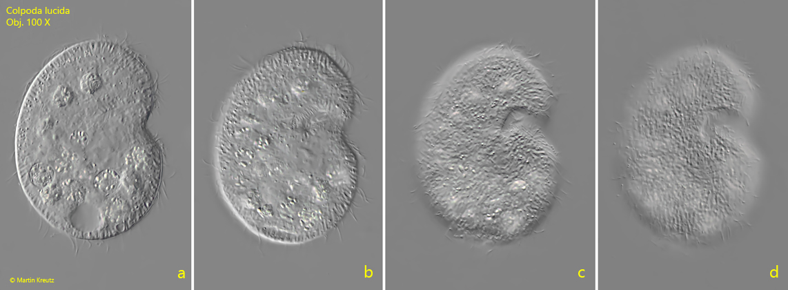
Fig. 1 a-d:Colpoda lucida. L = 82 µm. Different focal planes from right of a freely swimming specimen. Obj. 40 X.
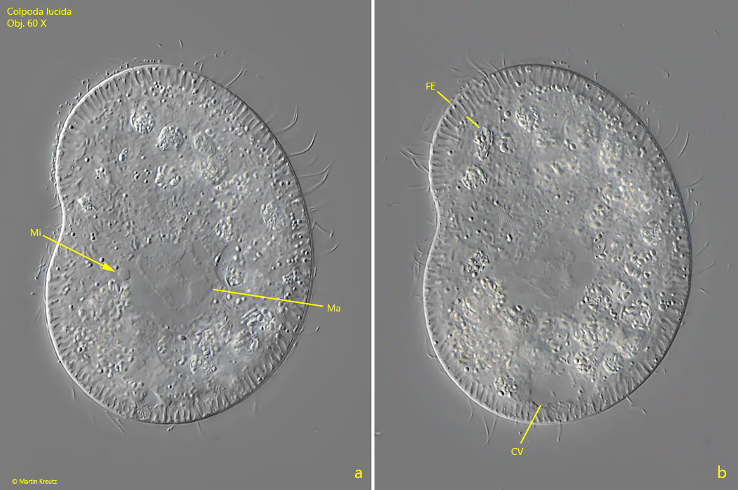
Fig. 2 a-b:Colpoda lucida. L = 90 µm. Two slightly different focal planes of a squashed specimen from left. Note the distinct fringe of extrusomes (FE). CV = contractile vacuole, Ma = macronucleus with reticulate nucleolus, Mi = micronucleus. Obj. 60 X.
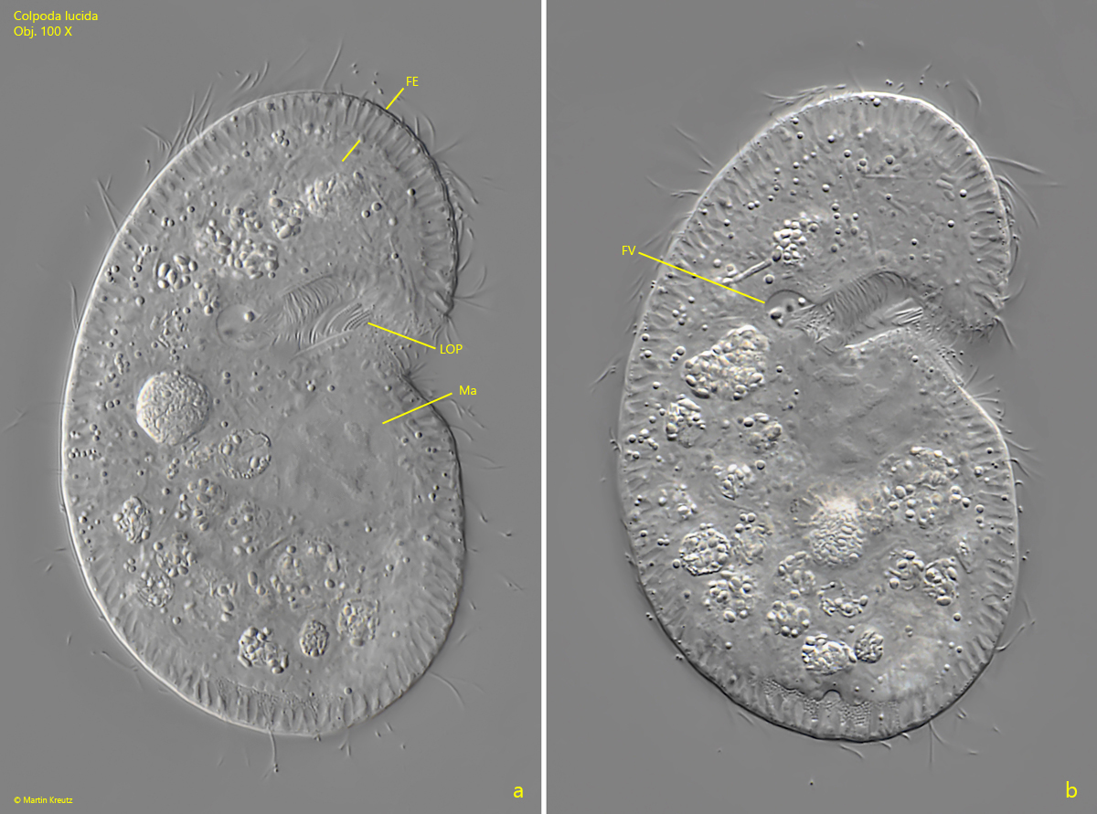
Fig. 3 a-b:Colpoda lucida. L = 88 µm. Two focal planes of a slightly squashed specimen from right. The fringe of extrusomes (FE) is 5 µm wide. At the end of the cytopharynx a food vacuole (FV) is being formed. LOP = left oral polykinetid, Ma = maronucleus. Obj. 100 X.
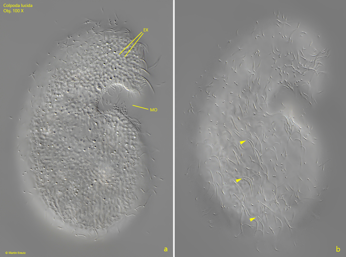
Fig. 4 a-b:Colpoda lucida. L = 88 µm. a) Focal plane on the layer of extrusomes (EX) which appear like blisters in DIC. b) focal plane on the somatic ciliation. Note the paired cilia (arrowheads) forming the kineties. Obj. 100 X.