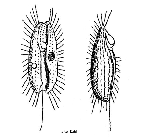
ventral side with a deep furrow running longitudinally
oral apparatus in the middle of the furrow
undulating membrane on left side of oral opening is pouch-shaped
macronucleus oval or elliposoid, on right side of body
one micronucleus adjacent to the macronucleus
one caudal cilium, inserted on right side of the postoral furrow
one contractile vacuole in the posterior third on the left side of the furrow
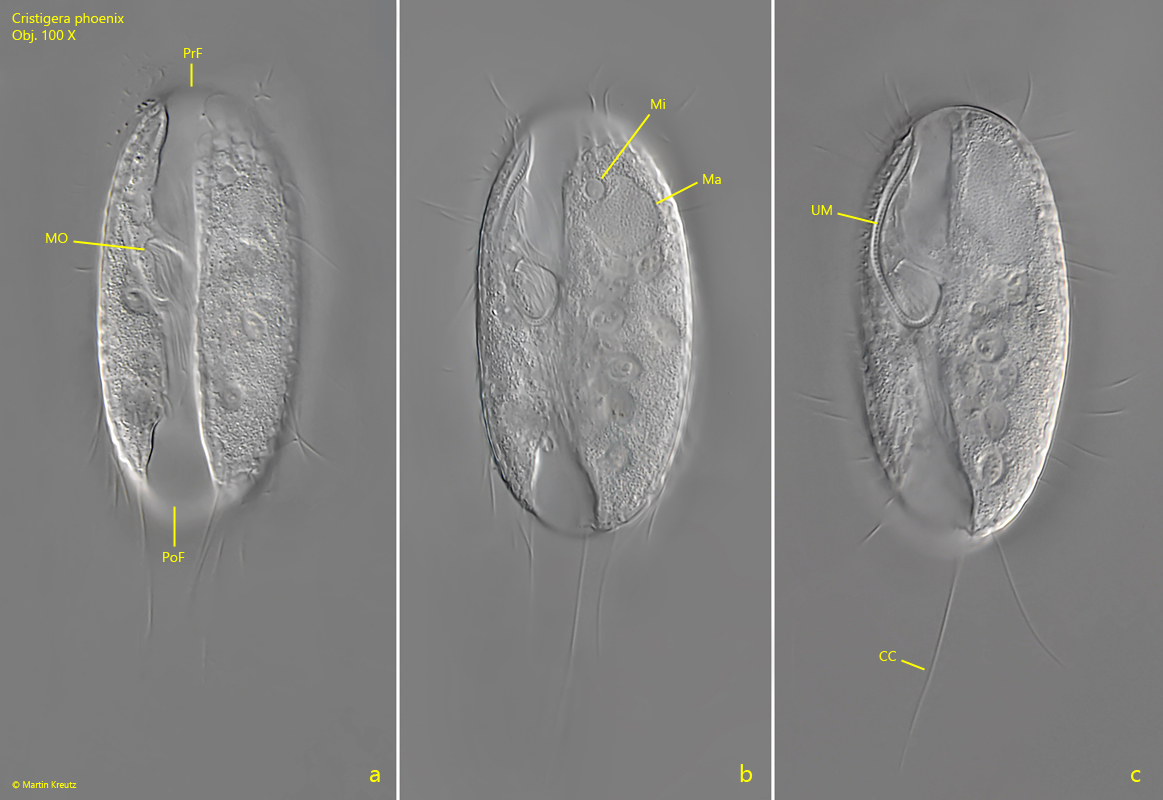
Cristigera phoenix
Due to the shape of Cristigera phoenix with a ventral furrow, Penard was reminded of a date palm kernel (Phoenix dactylifera) when he described the ciliate in 1922. That is where the species name derives from. I find Cristigera phoenix frequently and regularly especially in floating detritus flakes in old samples.
By the ventral furrow the species can be clearly identified as Cristigera phoenix. The similar species Cristigera minor is smaller and plumper. Cristigera penardi and Cristigera pleuronemoides have also a ventral furrow but the mid-body of these species is naked.
In a slightly squashed specimen I could observe that the cilia of the kineties are arranged in pairs (s. fig. 3 a) while Kahl drew them singly. The contractile vacuole was also shifted more to the posterior end in my specimens than Kahl drew it (s. drawing above). Possibly this is within the usual variability. Otherwise all features agree with the description and drawing of Kahl.
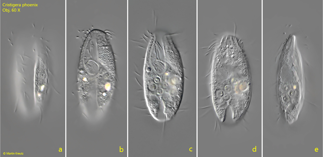
Fig. 1 a-e:Cristigera phoenix. L = 34 µm. Different focal planes of a freely swimming specimen from ventral (a–d) and laterally from right (e). Obj. 60 X.
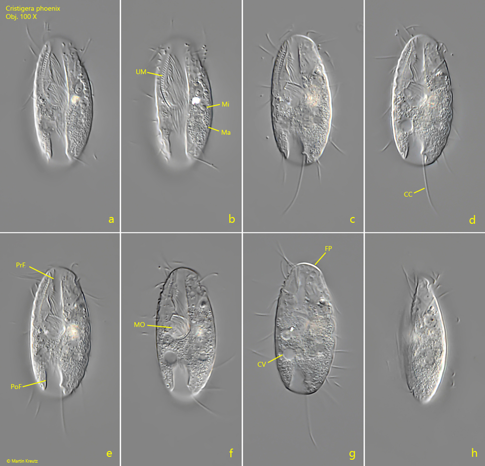
Fig. 2 a-h:Cristigera phoenix. L = 31 µm. A freely swimming specimen from ventral (a–g) and laterally from left (h). Note the ventral furrow divided into a praeoral (PrF) and postoral part (PoF) by the mouth opening (MO). The undulating membrane (UM) with long cilia is on the left side of the oral apparatus and the caudal cilium (CC) inserted at the right side of the postoral furrow (c, d). CV = contractile vacuole, FP = frontal plate, Ma = macronucleus, Mi = micronucleus. Obj. 100 X.
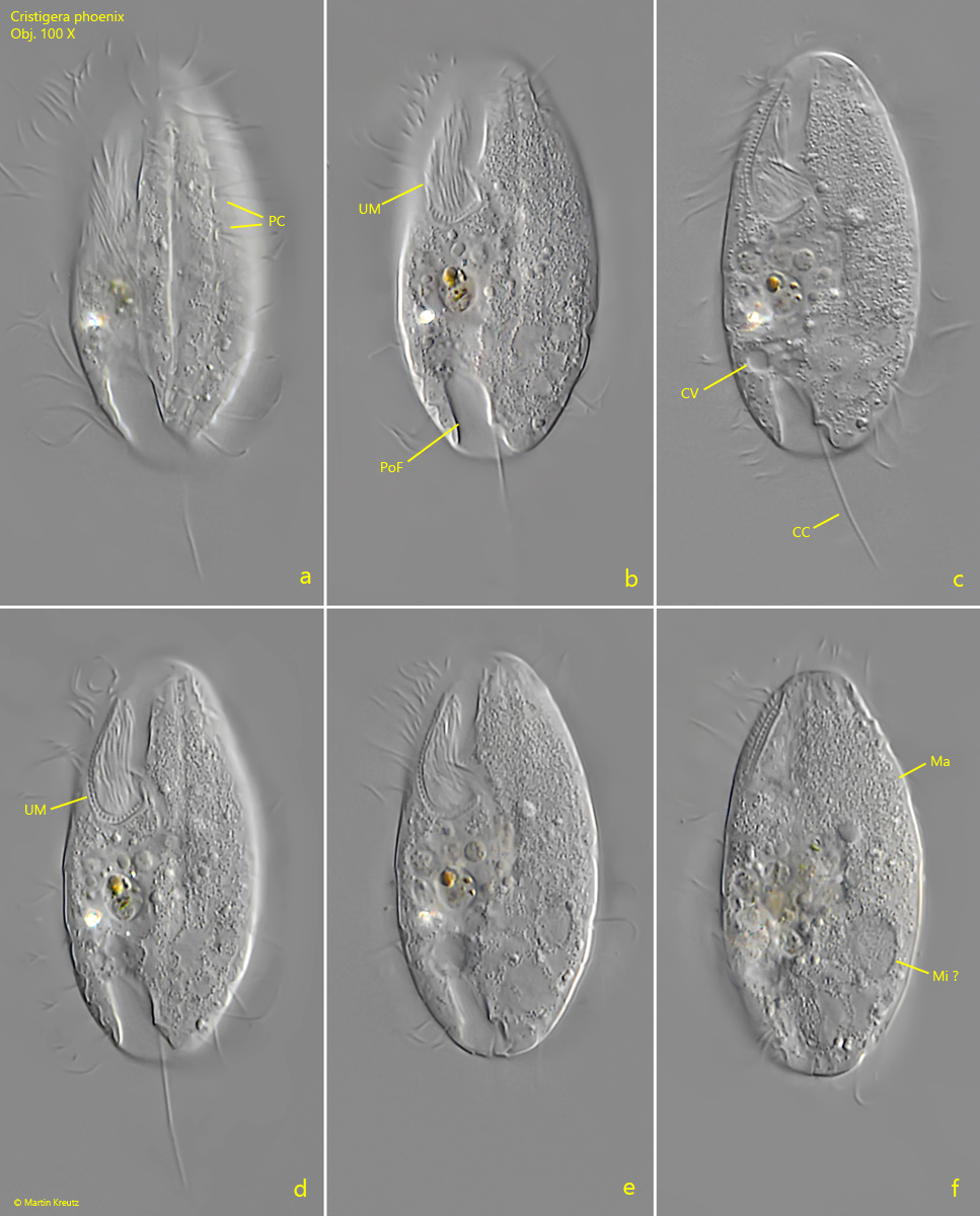
Fig. 3 a-f:Cristigera phoenix. L = 48 µm. A slightly squashed specimen from ventral. Note the paired cilia (PC, a) of the kineties. CC = caudal cilium, CV = contractile vacuole, Ma = macronucleus, Mi = probably the micronucleus, PoF = postoral furrow, UM = undulating membrane. Obj. 100 X.
Fig. 4 a-c:Cristigera phoenix. L = 58 µm. A freely swimming larger specimen. CC = caudal cilium, Ma = macronucleus, Mi = micronucleus, MO = mouth opening, PoF = postoral furrow, PrF = praeoral furrow, UM = undulating membrane. Obj. 100 X.