body long ellipsoidal with unciliated frontal plate
length 40–55 µm
oral apparatus small, subapical
globular macronucleus below cell equator
one micronucleus adjacent to macronucleus
extrusomes spindle-shaped, about 4 µm long
contractile vacuole in posterior fifth
cytoplasm filled with refractile granules, not ring-shaped
granules arranged in rows beneath pellicle, 8–9 rows per body side
one long caudal cilium
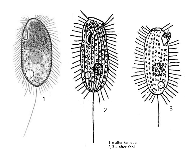
Dexiotricha elliptica
Dexiotricha elliptica was first described by Kahl in 1931 as Loxocephalus ellipticus. In 2014, Fan et al. transferred the species to the genus Dexiotricha on the basis of morphological investigations and renamed it Dexiotricha elliptica.
I find Dexiotricha elliptica quite frequently in the mud layer of decomposed leaves in Ulmisried and Purren pond. The ciliate is filled with granules, which are never ring-shaped in my specimens. Instead, the granules are mostly spherical or slightly asymmetrical in shape with a maximum diameter of 1.5 µm. Under the pellicle, the granules are arranged in longitudinal rows, as described by Kahl. IN my population the macronucleus was always located below the cell equator, and the contractile vacuole in the posterior fifth. I was also able to recognize the short canal between the contractile vacuole and the excretory porus on the ventral side, as drawn by Kahl (s. drawing 3 above).
Dexiotriche elliptica differs from the similar species Loxocephalus moniligranulatus and Dexiotricha granulosa mainly in the shape of the granules and the position of the contractile vacuole. In Loxocephalus moniligranulatus the contractile vacuole is located in the middle of the body and in Dexiotricha granulosa the granules are ring-shaped and the contractile vacuole is also located in the middle of the body.
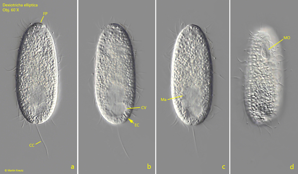
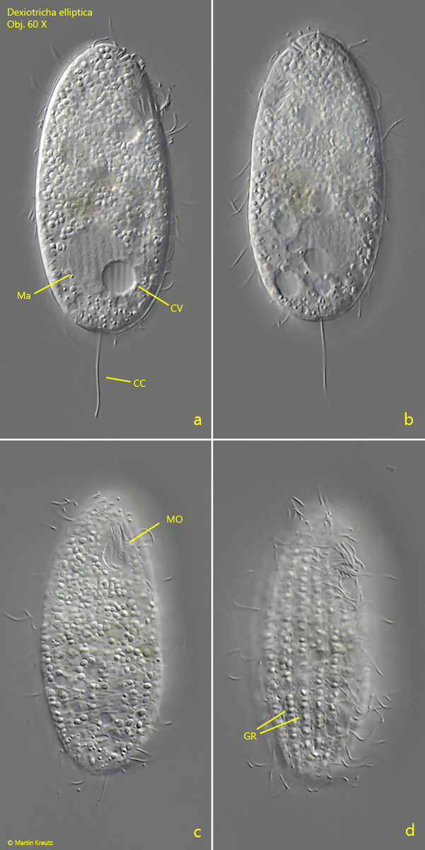
Fig. 1 a-d:Dexiotricha elliptica. L = 52 µm. A freely swimming specimen. Note the short excretion canal (EC) of the contractile vacuole. CC = caudal cilium, CV = contractile vacuole, FP =frontal plate, Ma = macronucleus, MO = mouth opening. Obj. 60 X.
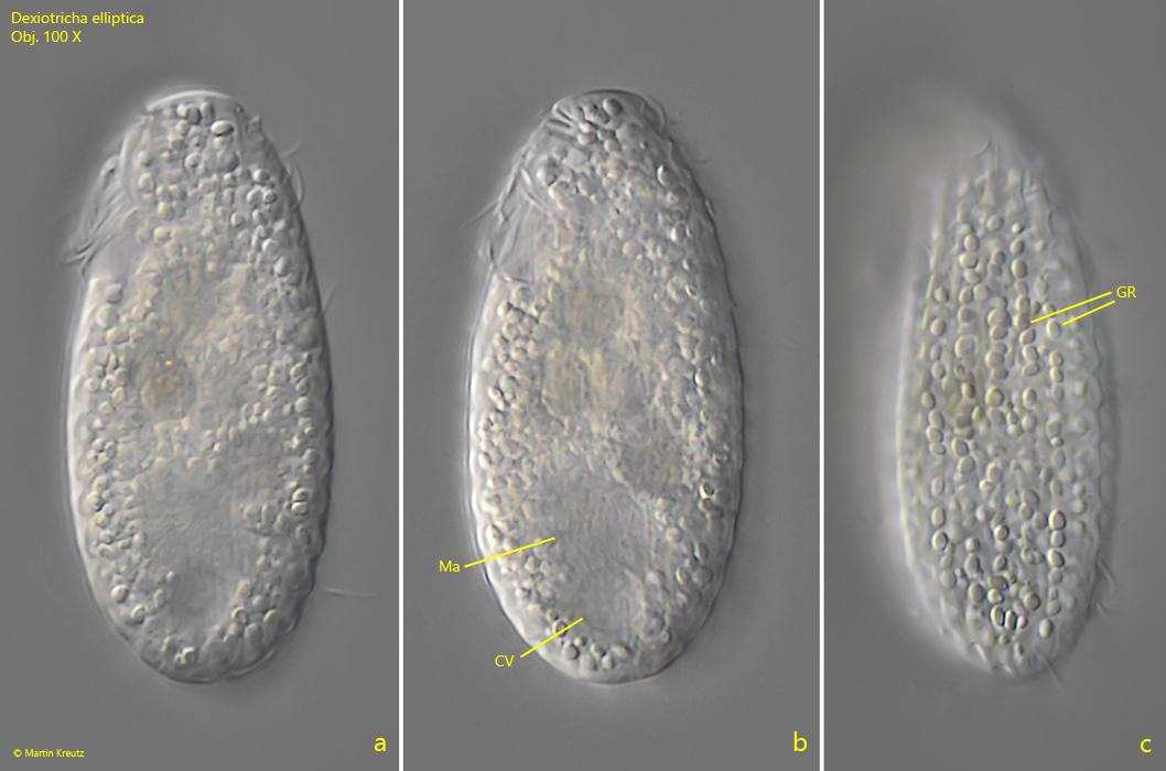
Fig. 2 a-c:Dexiotricha elliptica. L = 52 µm. The slightly squashed specimen as shown in fig. 1 a-d. Note the granula (GR) arranged in longitudinal rows beneath the pellice. CV = contractile vacuole, Ma = macronucleus. Obj. 100 X.
Fig. 3 a-d:Dexiotricha elliptica. L = 59 µm. Different focal planes of a second, slightly squashed specimen. CC = caudal cilium, CV = contractile vacuole, GR = granules, Ma = macronucleus, MO = mouth opening. Obj. 60 X.
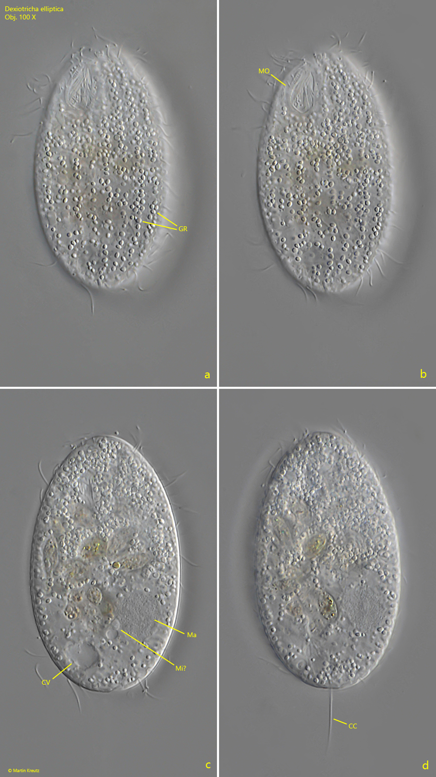
Fig. 4 a-d:Dexiotricha elliptica. Different focal planes of a strongly squashed squashed specimen. CC = caudal cilium, CV = contractile vacuole, GR = granules, Ma = macronucleus, Mi? = probably the micronucleus, MO = mouth opening. Obj. 100 X.
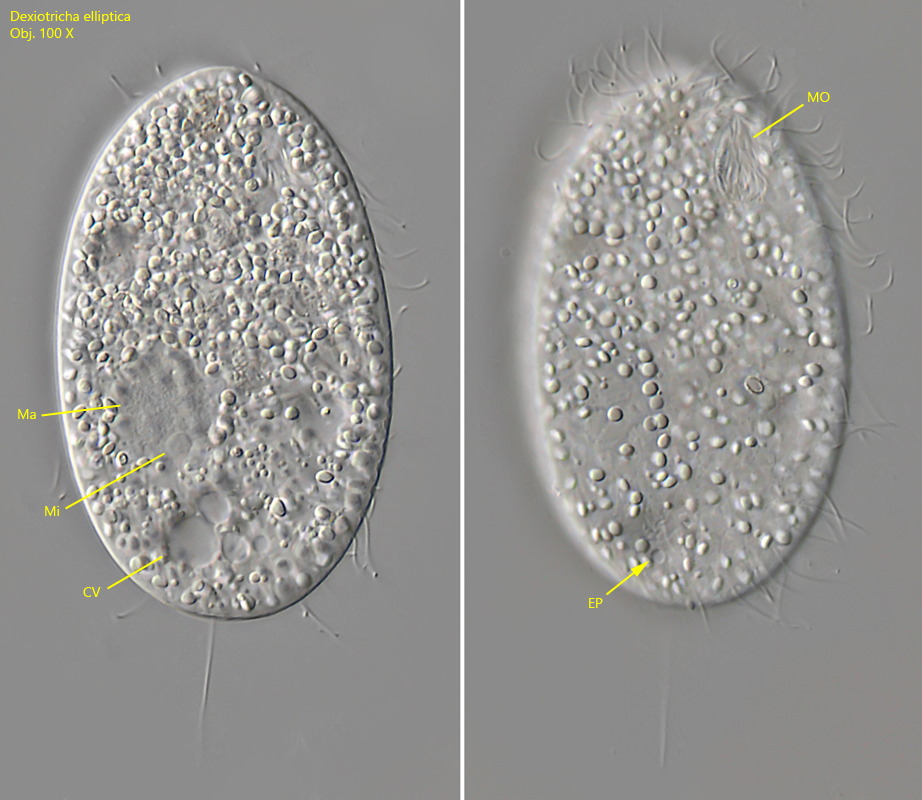
Fig. 5 a-b:Dexiotricha elliptica. A second strongly squashed squashed specimen. Note the excretion pore (EP) of the contractile vacuole. CV = contractile vacuole, Ma = macronucleus, Mi = micronucleus, MO = mouth opening. Obj. 100 X.