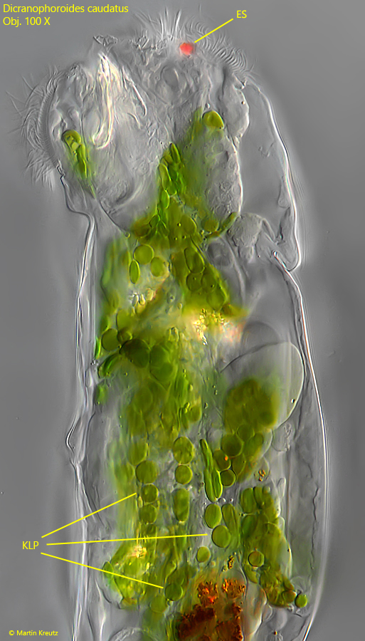
Dicranophoroides caudatus is very common in my localities Simmelried and Ulmisried and is always found in detritus. There it feeds on Euglenophyceae, like Euglena, Lepocinclis or Phacus (s. fig. 4). The species is very easy to identify by its green coloration. This is caused by so-called kleptoplasts. These are “stolen” chloroplasts that come from the prey organisms (s. fig. 5). The chlorplasts are not digested like the rest of the prey, but are stored intracellularly while retaining their full function. The selection of chloroplasts appears to be selective. I have only been able to find disc-shaped chloroplasts in Dicranophoroides caudatus, such as those found in Lepocinclis acus. In starving specimens the number of kleptoplasts can be strongly reduced up to almost colorless specimens.
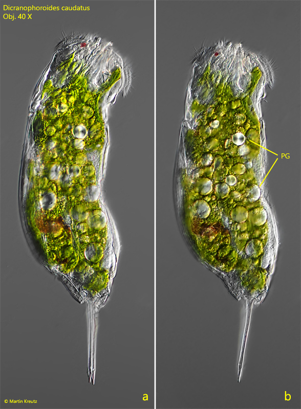
Fig. 1 a-b: Dicranophoroides caudatus. L = 240 µm. A freely swimming, well fed specimen in lateral view from right. The stomach is complety filled with disc-shaped paramylon grains (PG), most likely from Phacus species. Obj. 40 X.
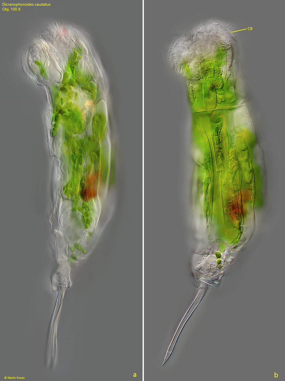
Fig. 2 a-b: Dicranophoroides caudatus. L = 264 µm. A slightly squashed specimen in detail. CB = circumapical band of cilia. Obj. 100 X.
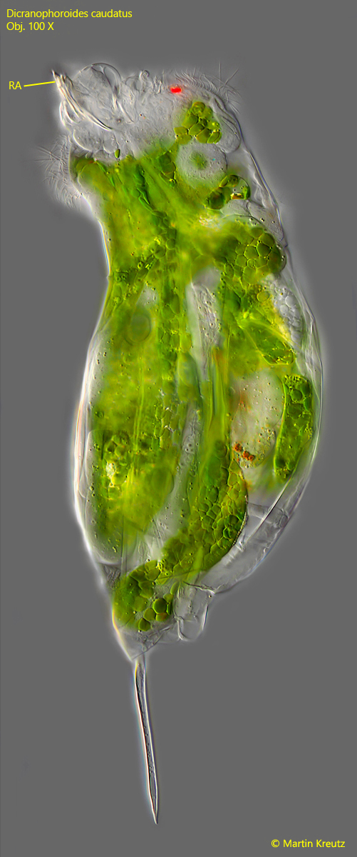
Fig. 3: Dicranophoroides caudatus. L = 264 µm. In this squashed specimen, the rami protrude from the mouth with the teeth at the distal ends. RA = rami with distal teeth. Obj. 100 X.
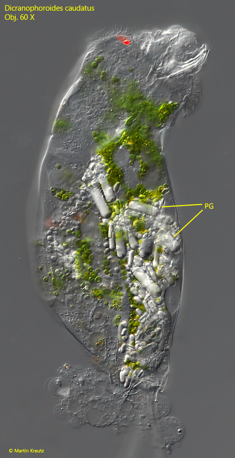
Fig. 4: Dicranophoroides caudatus. In this squashed specimen the paramylon grains (PG) of ingested Euglenophyceae (likely Lepocinclis acus) are visible. Obj. 60 X.
Fig. 5: Dicranophoroides caudatus. The green coloration is caused by chloroplasts derived from phagocytosed Euglenophyceae (e.g. Euglena, Lepocinclis or Phacus). The chloroplasts of the prey are not digested but are stored intracellularly and maintained in full function. They are then referred to as kleptoplasts (KLP). ES = eye spot. Obj. 100 X.