anterior end bluntly rounded, posterior end bluntly pointed or rounded
length 20–33 µm
two band-shaped chloroplasts with slightly lobed margins
each chloroplast contains a double layer pyrenoid
paramylon grains small, oval, ovoid or short rods
eyespot medium-sized
nucleus located posterior
flagellum up to twice body length
pellicle faintly striated in counterclockwise direction
Euglena agilis
Euglena agilis is a very common, globally distributed species and has also been described independently as Euglena pisciformis (Klebs, 1883) and Euglena nana (Johnson, 1944). The name Euglena agilis, first given by Carter, has prevailed.
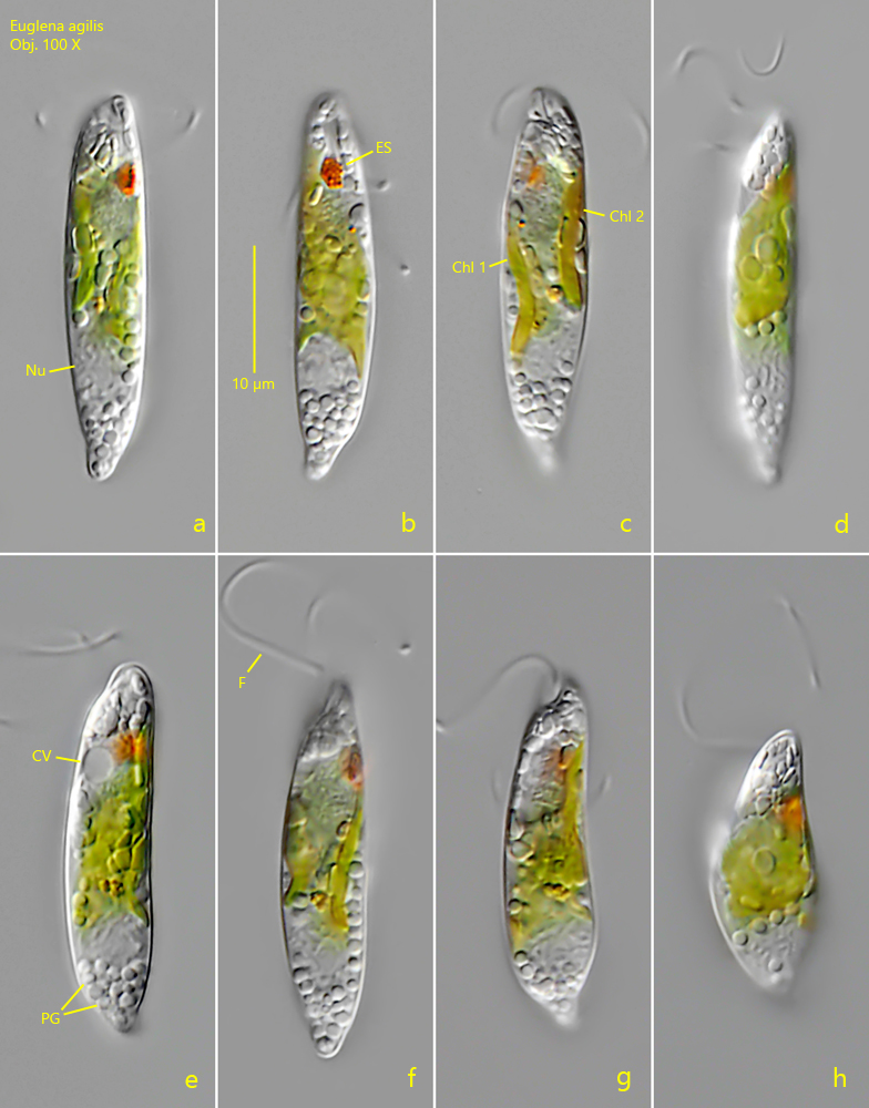
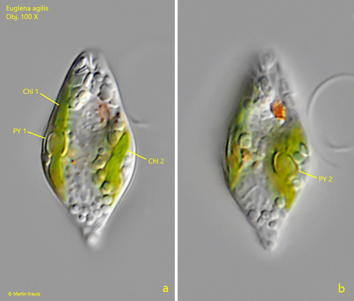
I found large numbers of Euglena agilis in August 2023 in a heavily eutrophic pond in the Wollmatinger industrial area. The fast-swimming cells appear spindle-shaped at low magnification. At higher magnification, however, the body appears almost cylindrical and parallel-sided. The anterior end is rounded and in my population the posterior end was somewhat cone-shaped and rounded (s. fig. 1a). The most important feature for the identification of Euglena agilis are the two ribbon-shaped chloroplasts (s. fig. 1 c and 2 a). In each of of the chloroplasts a double layer pyrenoid is located (s. fig. 2 a-b). These double layer pyrenoids are also the distinguishing feature from the similar species Euglena van-goori, which also has two chloroplasts, but without pyrenoids.
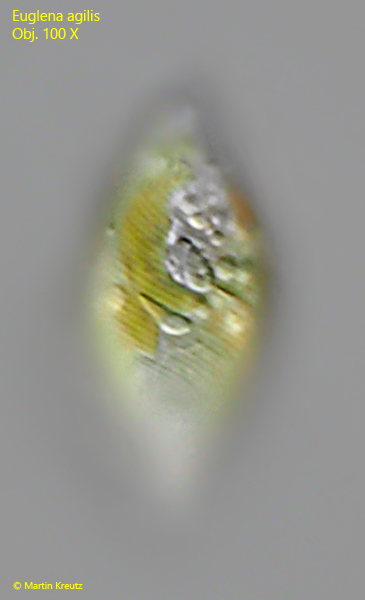
The nucleus is found near the posterior end and the paramylon grains in my population were small and broadly oval or spherical. In many specimens an I could observe an accumulation of paramylon in the posterior end (s. fig. 1 e). The striation of the pellicle is faint and runs in counterclockwise direction (s. fig. 3). The flagellum of the specimens in my population was almost twice as long as the cell.
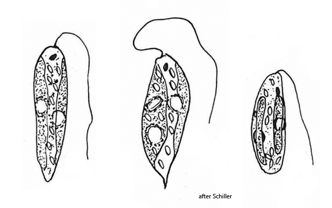
Schilling (1956) studied the population of Euglena agilis from 1950-1955 in Lake Neusiedl (Austria) in detail. He points out the high variability of the cell shapes. He found forms with pointed posterior ends or almost oval specimens (s. drawings above). The two ribbon-shaped chloroplasts, each with a double layer pyrenoid, proved to be an essential, constant characteristic.
Fig. 1 a-h:Euglena agilis. L = 32 µm. A freely swimming specimen. Note the two, band-shaped chloroplasts (Chl 1 + Chl 2, c). Obj. 100 X.
Fig. 2 a-b:Euglena agilis. A slightly squashed specimen with focal planes on the double layer pyrenoids (PY 1, PY 2) in each of the two chloroplasts (Chl 1, Chl 2). Obj. 100 X.
Fig. 3: Euglena agilis. Focal plane on the faint striation of the pellicle running in counterclockwise direction. Obj. 100 X.