Goniomonas 1
Most likely ID: Goniomonas nov. spec.
Synonym: n.a
Sampling location: Simmelried
Phylogenetic tree: n.a.
Diagnosis:
- cell ovate, posteriorly rounded, obliquely truncate anteriorly
- laterally flattened
- dorsally (position of flagella) rounded, ventrally (position of contractile vacuole) a clear indentation
- ventral margin folded over to the right side at the level of the cell center
- length 22–24 µm, width 16–18 µm
- two flagella of equal length, half length of body
- 7–9 ejectisomes arranged parallel to anterior margin
- a cluster of 5–8 irregularly arranged ejecticomes below the position where flagella arise
- one contractile vacuole in opposite position to the flagella
- periplast with each 4 dotted stripes on each side
- each two stripes are joining at posterior end
- nucleus near mid-body at dorsal side
No drawings from previous authors available.
In January 2023 I found a Goniomonas species in the Simmelried, which is clearly different from the previously described species Goniomonas truncata, Goniomonas pacifica and Goniomonas amphinema (Larsen & Patterson 1990, s. Literature). The cells were 22–24 µm long, which is twice as large as the length of Goniomonas truncata (5–12 µm), Goniomonas pacifica (8–10 µm) and Goniomonas amphinema (5-8 µm). In addition, there are other features that are not found in the previously described species. At the dorsal end of the row of 7–9 parallel arranged ejectisomes there is another disordered cluster 0f 5–8 ejectisomes (s. figs. 1b and 2a). The anterior margin is folded over to the right side in the middle, creating a ventral indentation (s. figs. 1c and 2c). On each side of the body there are 4 dotted stripes (s. figs. 1d and 2c). These do not run slightly oblique to the posterior end, as in Goniomonas truncata, for example, but in slightly angled lines (s. fig. 1d). Near the posterior end, two of the stripes join each other (s. fig. 2c). The cell shape is generally oval, but with some straight facets, especially in the anterior half. The two flagella are of equal length and with 11–12 µm about half as long as the body (s. fig. 2c). I found this species on coverslips floating on samples with decomposing plant material from the Simmelried.
Because of the large differences to the previously described Goniomonas species and the lack of alternatives, I consider this to be a new, not yet described species (Goniomonas nov. spec.).
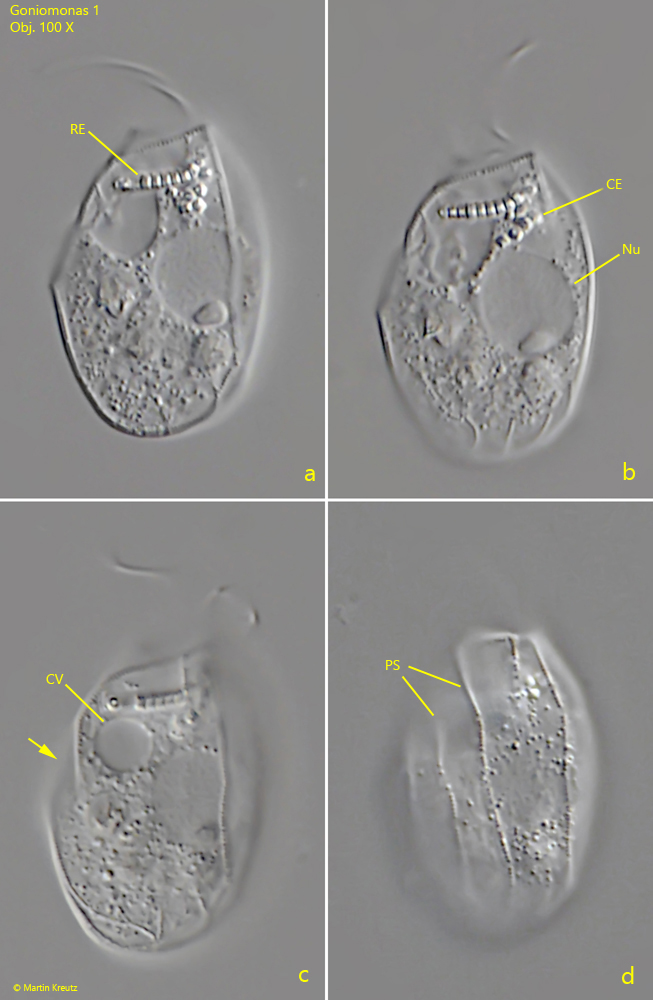
Fig. 1 a-d: Goniomonas 1. L = 23.3 µm. Different focal planes from the left side. Note the cluster of irregularly arranged ejectisomes (CE) below the row of parallel arranged ejectisomes (RE) as well as the ventral margin folded to the right side (arrow). CV = contractile vacuole, Nu = nucleus, PS = periplast striation (dotted). Obj. 100 X
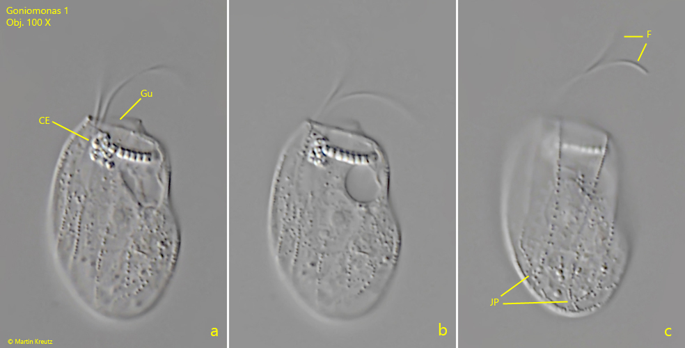
Fig. 2 a-c: Goniomonas 1. L = 22.3 µm. Different focal planes from the right side of a second specimen. Note that each two stripes of the periplast striation joining near the posterior end (JP). The flagella (F) have a equal length of 11–12 µm. CE = cluster of irregularly arranged ejectisomes, Gu = gullet. Obj. 100 X