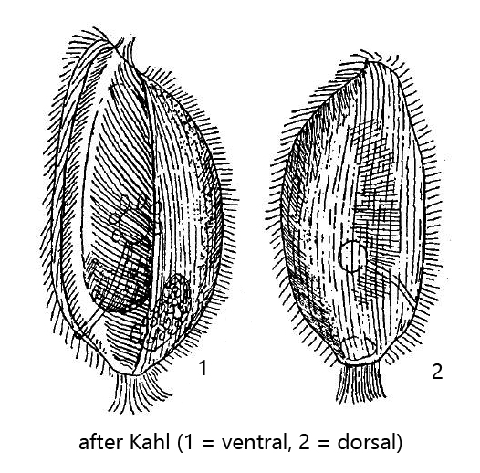
very large oral apparatus reaching down to the posterior end
left side of oral apparatus with adoral membranelle of cilia glued together
right side of oral apparatus with an undulating membrane (hard to see)
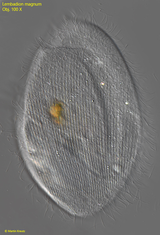
The pellicle shows a longitudinal striation on the anterior and posterior side
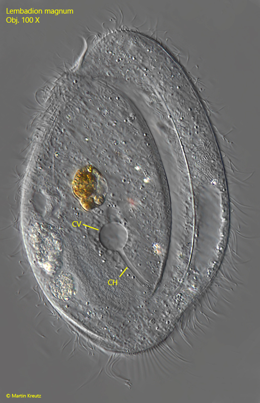
contractile vacuole dorsally in mid-body, connected via a channel with the ventral excretion porus
macronucleus kidney-shaped in the posterior third with an adjacent spherical micronucleus
posterior end with a tuft of cilia
Lembadion magnum
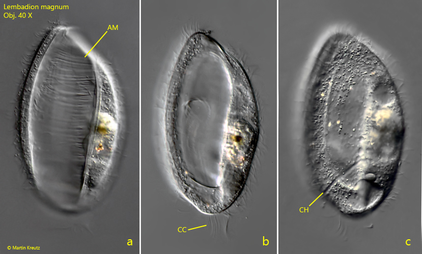
Lembadion magnum is present in all of my sites. The species does not seem to have very specific requirements for environmental conditions. One recognizes Lembadion magnum immediately by the huge adoral membranelle, which extends over the entire body length (s. figs. 1 a, 2 a and 3 a). This is also a feature distinguishing Lembadion magnum safely from Lembadion lucens and Lembadion bullinum, in which the adoral membranelle extends only to the posterior third of the body. In addition, Lembadion magnum is larger and the pellicle is not characterised by a pattern of regularely arranged squares, but has a longitudinal striation (s. fig. 4).
Fig. 1 a-c: Lembadion magnum. L = 124 µm. Ventral view of a freely swimming specimen in three focal planes. AM = adorale membranelle, CC = tuft of caudal cilia, CH = channel between contractile vacuole and ventral excretion porus. Obj. 40 X.
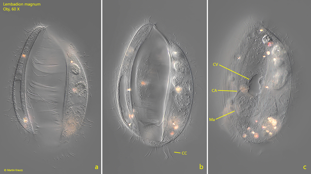
Fig. 2 a-c: Lembadion magnum. L = 110 µm. Ventral view of a second freely swimming specimen in three focal planes. CA = channel between contractile vacuole and ventral excretion porus, CC = tuft of caudal cilia, CV = contractile vacuole, Ma = macronucleus. Obj. 60 X.
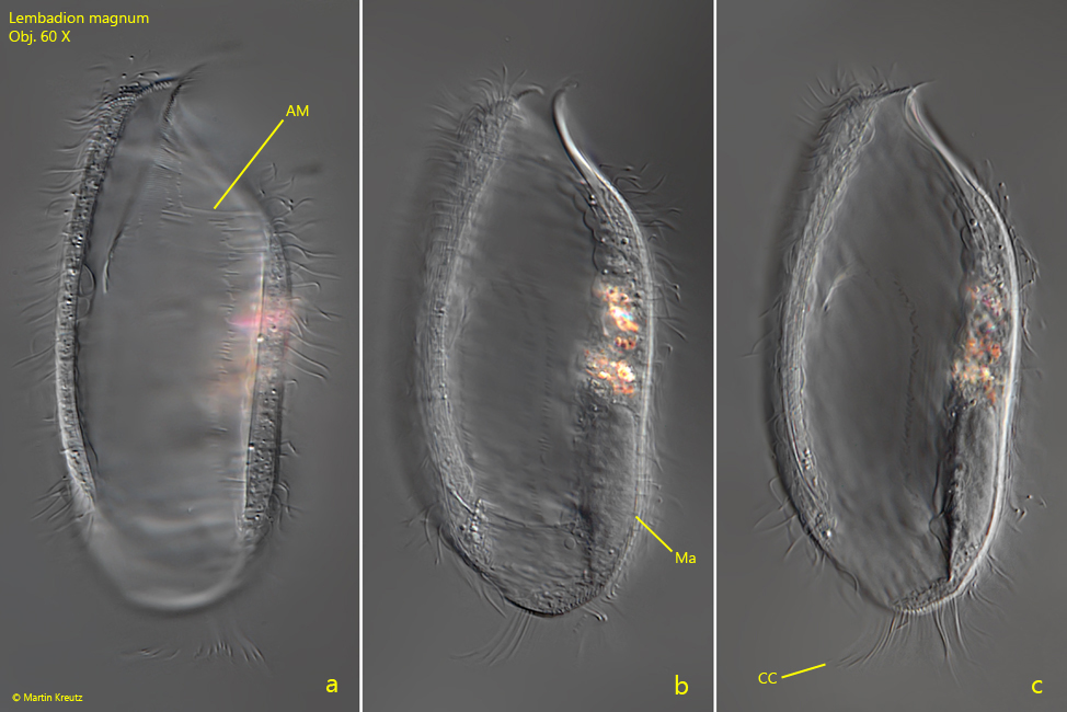
Fig. 3 a-c: Lembadion magnum. L = 160 µm. Ventral view of a third freely swimming specimen in three focal planes. AM = adorale membranelle, CC = tuft of caudal cilia, Ma = macronucleus. Obj. 100 X.
Fig. 4: Lembadion magnum. Dorsal view of a slightly squashed specimen with the longitudinal striation of the pellicle. Obj. 100 X.
Fig. 5: Lembadion magnum. Dorsal view of a slightly squashed specimen with focal plane on the channel (CH) connecting the contractile vacuole (CV) with the ventrally located excretion porus. Obj. 100 X.