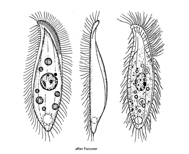
body elongated and flattened, ventral side convex, dorsal side straight or slightly sigmoidal
right side flat with 3–4 rows of cilia
left side convex with 3–4 rows of bristles
dorsal brush a third of body length
length 30–50 µm, width 10–15 µm
one ellipsoid maronucleus to which one spherical micronucleus is closely attached
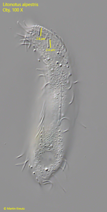
rod-shaped extrusomes 2–3 µm long, arranged along oral cleft
contractile vacuole subterminal
Litonotus alpestris
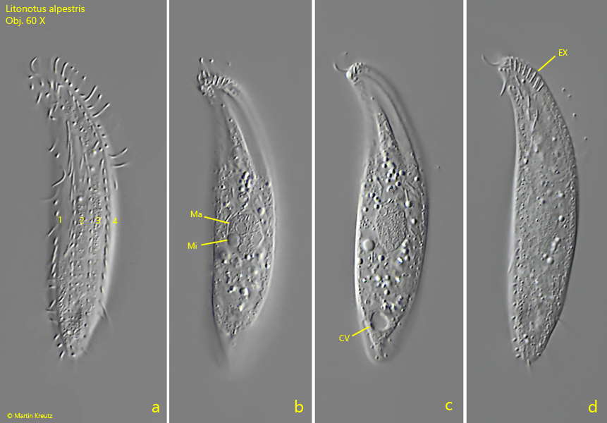
Litonotus alpestris was only discovered in 1978 and described by Foissner. This is actually surprising because this small Litonotus species is very common. However, it is difficult to detect in fresh samples because often the specimens “hide” in detritus flocs. However, they can be easily “extracted” on “floating coverslips“, where Litonotus alpestris likes to settle. The specimens settling there always point with the right, ciliated side to the observer, with which they slide along the coverslip. Therefore all images shown below are taken from the right side.
I have also made images of Litonotus alpestris in brightfield illumination (s. fig. 1 a-d). These images show the difference to the DIC images below. For the early observers (i.e. before DIC and phase contrast) it must have been a challenge to capture and describe such transparent objects.
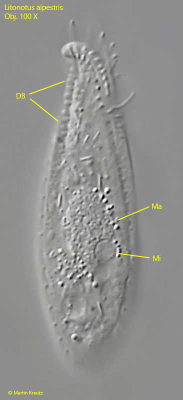
Litonotus alpestris differs from most other Litonotus species in the nuclear apparatus. The species has only one macronucleus with an attached micronucleus (s. figs. 2 a and 2 b) and thus differs from the similar species Litonotus lamella, which has two macronuclei.
Fig. 1 a-d:Litonotus alpestris. L = 56 µm. A freely gliding specimen in brightfield illumination from right. Although the specimen is highly transparent the macronucleus (Ma) and the attached micronucleus (Mi) are visible. CV = contractile vacuole. Obj. 100 X.
Fig. 2 a-d:Litonotus alpestris. L = 55 µm. A second freely gliding specimen from right. Note the 4 rows of cilia of the right side (a, 1–4). CV = contractile vacuole, EX = extrusomes, Ma = macronucleus, Mi = micronucleus. Obj. 60 X.
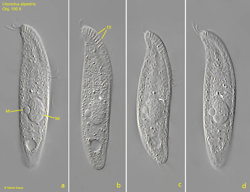
Fig. 3 a-d:Litonotus alpestris. L = 55 µm. A third freely gliding specimen from right. EX = extrusomes, Ma = macronucleus, Mi = micronucleus. Obj. 100 X.
Fig. 4:Litonotus alpestris. L = 39 µm. Focal plane on the dorsal brush (DB). Ma = macronucleus, Mi = micronucleus. Obj. 100 X.
Fig. 5:Litonotus alpestris. The extrusomes are rod-shaped and 2.4–2.8 µm long. Obj. 100 X.