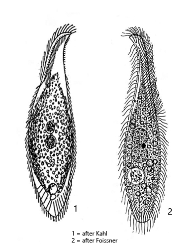
body slenderly lanceolate and flattened, ventral side convex, dorsal side straight or slightly curved
posterior end broadly rounded
right side flat with 4–7 rows of cilia
left side convex with 4 rows of bristles
dorsal brush a third of body length
length 50–100 µm, width 10–25 µm
extrusomes rod-shaped and slightly curved, 5–7 µm long
extrusomes arranged along oral cleft and at posterior end
two closely spaced, spherical macronuclei
one spherical micronucleus between the macronuclei
contractile subterminal
Litonotus lamella
I find Litonotus lamella in almost all of my sampling sites. The species is very common. For identification the shape of the extrusomes and their distribution is important (at the oral cleft as well as in the posterior end) as well as the number of macronuclei. It must be two, between which the small micronucleus is located. Litonotus lamella can be easily confused with Litonotus alpestris(has only one macronucleus) and with Litonotus crystallinus (has several longitudinal ribs on the left side).
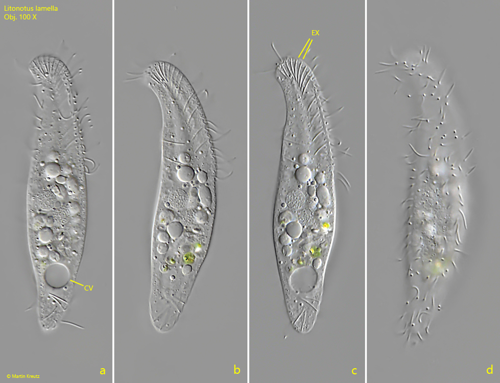
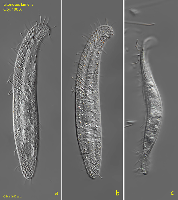
In my experience, Litonotus lamella is best observed on the floating coverslip, on which the ciliate likes to settle. Since it slides along the coverslip with the ciliated (right) side, it can almost always be seen from the right (s. fig. 1 a-d). To see it from the left (s. figs. 2 a-c and fig. 3 a-b), the sample must be pipetted on the slide and than you need to wait some minutes before placing the coverslip. During this time, all specimens in the sample will turn with the right side towards the slide to glide on it.
Fig. 1 a-d:Litonotus lamella. L = 66 µm. A freely gliding specimen from the right side. CV = contractile vacuole, EX = extrusomes. Obj. 100 X.
Fig. 2 a-c:Litonotus lamella. L = 85 µm. A second, freely gliding specimen from the left side (a, b) and from ventral (c). Obj. 100 X.
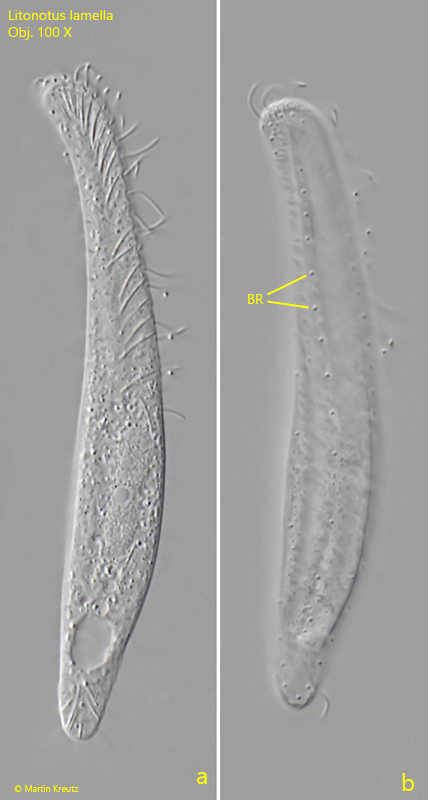
Fig. 3 a-b:Litonotus lamella. L = 82 µm. A third specimen from the left side. Note the rows of short bristles (BR) of the left side. Obj. 100 X.
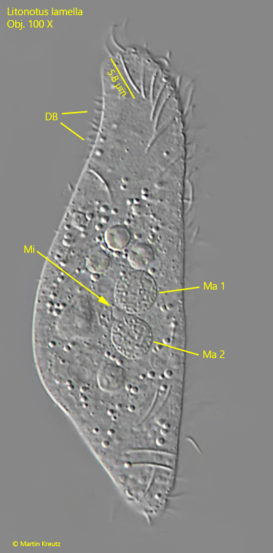
Fig. 4:Litonotus lamella. L = A strongly squashed specimen. Note the spherical micronucleus (Mi) between the macronuclei (Ma 1, Ma 2). DB = dorsal brush. Obj. 100 X.
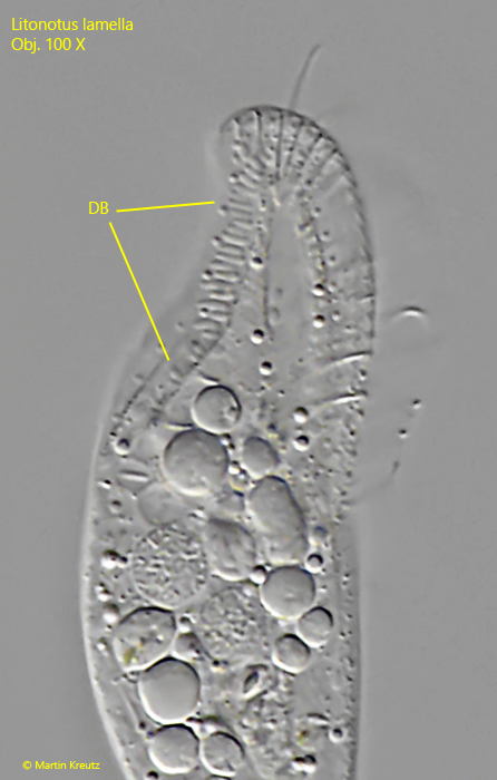
Fig. 5:Litonotus lamella. The dorsal brush (DB) in detail. Obj. 100 X.