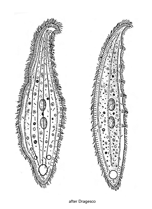
body lanceolate or knife-shaped, laterally flattened
length 75–300 µm (commonly about 200 µm)
ventrally broad seam of extrusomes
dorsally warts with bundles of extrusomes
right side ciliated, left side curved shaped and naked
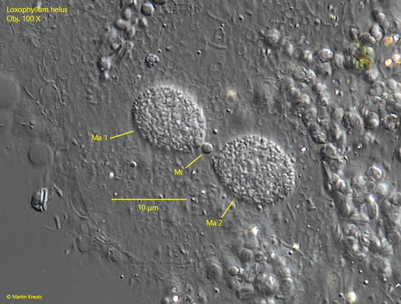
two spherical or ellipsoid macronuclei with a spherical or ellipsoid micronucleus between them
contractile vacuole subterminal
3 types of extrusomes, the largest about 8–10 long and slightly curved
terminal scales with keel at posterior end
Loxophyllum helus
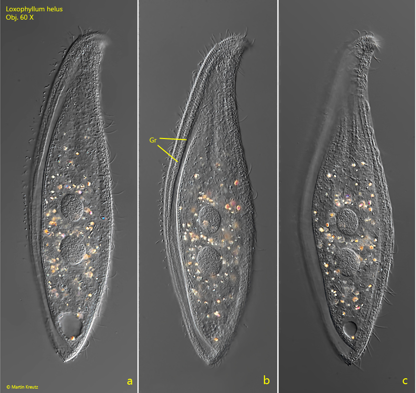
I find Loxophyllum helus rarely but regularly in the mud layer of several of my sites. This ciliate is easily identified because it has a distinct groove on the left side of the body that runs parallel to the body outline (s. fig. 1b). This is caused by the convex left side of the body, which is naked. In addition, Loxophyllum helus has a ventral seam of densely packed extrusomes that begins at the apical end, encircles the posterior end, and ends shortly thereafter on the dorsal side (s. fig. 1c). The dorsal side is covered with warts containing bundles of extrusomes, as is also the case with Loxophyllum meleagris. However, Loxophyllum helus also differs from this species in the nuclear apparatus, which consists of two macronuclei with a small spherical or oval micronucleus between them (s. figs. 5b and 8). Loxophyllum helus feeds mainly on small ciliates.
Fig. 1 a-c:Loxophyllum helus. L = 186 µm. A freely swimming specimen from left. Note the groove (Gr) caused by the convex shaped left side. Obj. 60 X.
Fig. 2 a-b:Loxophyllum helus. L = 214 µm. A second freely swimming specimen from right. Obj. 60 X.
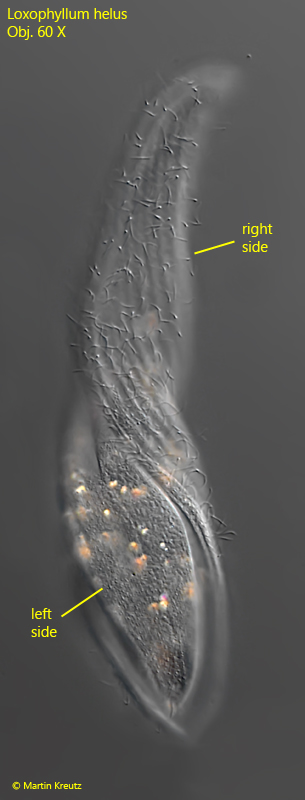
Fig. 3:Loxophyllum helus. On this twisted specimen the naked left side and the ciliated right side is visible. Obj. 60 X.
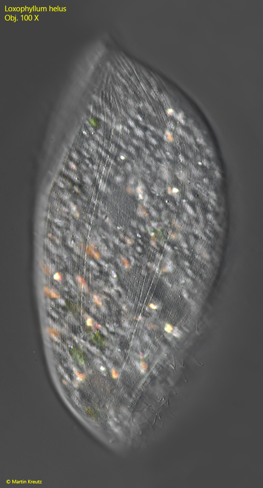
Fig. 4:Loxophyllum helus. The naked left side with some fine grooves. Obj. 100 X.
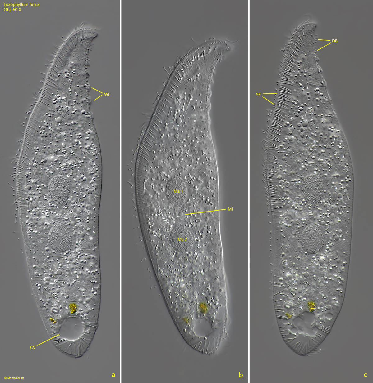
Fig. 5 a-c:Loxophyllum helus. A slightly squashed specimen from left. CV = contractile vacuole, DB = dorsal brush; Ma 1, Ma 2 = macronuclei; Mi = micronuclus; SE = ventral seam of extrusomes; WE = dorsal warts with bundles of extrusomes. Obj. 60 X.
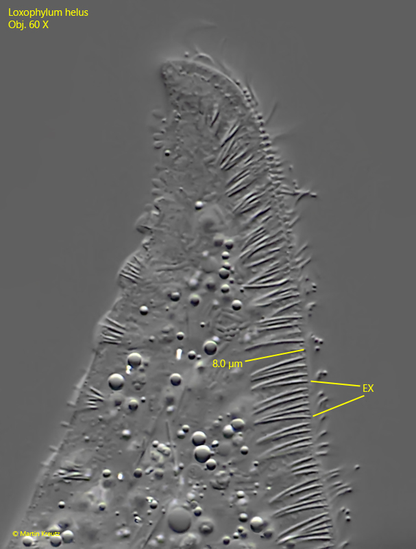
Fig. 6:Loxophyllum helus. The slightly curved extrusomes are 8 – 10 µm long. Obj. 60 X.
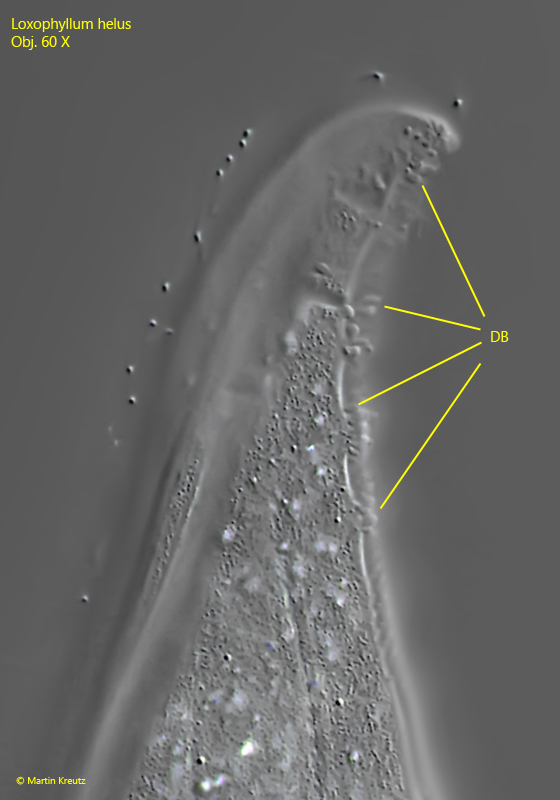
Fig. 7:Loxophyllum helus. Part of the dorsal brush, which consists of short, club-shaped bristles. Obj. 60 X.
Fig. 8:Loxophyllum helus. The two macronuclei (Ma 1, Ma 2) and the small, spherical micronucleus (Mi) between them. Obj. 100 X.