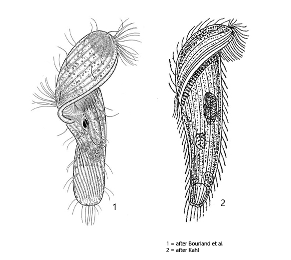
body oblong, S-shaped, tapered to the posterior end
length 90–160 µm
longitudinal rows of cilia, uniform ciliation, caudal cilia slightly elongated
acculumation of refractive granules in apical dome
perizonal stripe include 5 ciliary rows
adoral zone slightly spiralized, reaches mid-body
macronucleus oval or ellipsoidal
spherical micronucleus in depression of macronucleus
symbiotic bacteria in cytoplasm
contractile vacuole terminal
Metopus es
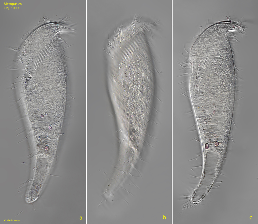
Metopus es, together with Metopus nasutus, is one of the most common metopid ciliates in my sampling locations. The species is easily recognized by its sigmoid shape and the size > 100 µm, although the sigmoid shape may not always be as pronounced as the specimen shown in fig. 1 a-c. Often the specimens are brownish in color, with a distinct dark brown spot in the apical dome caused by an accumulation of highly refractive granules (s. fig. 3). The adoral zone encircles the ventral side and terminates on the left side, so that the ciliate has to be slightly turned to see it (s. fig. 2 b). Metopus es swims quite fast, but can be easily fixed by coverslip pressure because the species is not as coverslip sensitive as many other metopid ciliates. In my population, specimens were almost always longer than 140 µm and often quite transparent. In the posterior half of the body, I was able to identify canals that opened into the contractile vacuole (s. fig. 3), which I believe to be auxiliary canals that have not been previously described for Metopus es. In 2016, a redescription of Metopus es was made by Bourland et al. But also by these authors no auxiliary channels of the contractile vacuole are mentioned. Further investigations must show whether this is a constant feature.
Fig. 1 a-c:Metopus es. L = 163. Three focal planes from ventral of a freely swimming specimen. Note the S-shape of the body. Obj. 100 X.
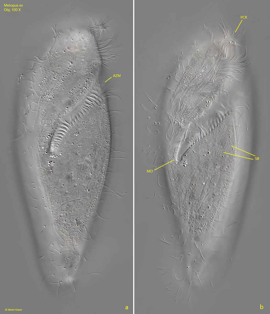
Fig. 2 a-b:Metopus es. L = 140 µm. Two focal planes from left of a slightly squashed specimen. AZM = adoral zone of membranelles, MO = mouth opening, PCR = perizonal ciliary rows, SB = symbiotic bacteria in the cytoplasm. Obj. 100 X.
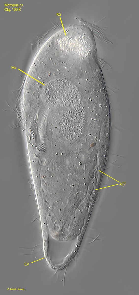
Fig. 3:Metopus es. L = 140 µm. The stronger squashed specimen shown in fig. 2 a-b with focal plane on the macronucleus (Ma). In the posterior half fine canals can be seen. They open into the contractile vacuole and are probably auxiliary channels (AC). CV = contractile vacuole, RG = accumulation of refractive granules in the apical dome. Obj. 100 X.