body long oval, tapered towards posterior end, posterior end truncated
length 60 – 90 µm, rarely larger
apical dome overhangs left side
adoral zone reaches almost mid-body
stripes widely ribbed
caudal cilia arise around the terminal pole
terminal pole free of caudal cilia
contractile vacuole terminal
excretion porus terminal
Metopus setifer
I rarely find Metopus setifer in the Simmelried. It is easily recognized by the long caudal cilia, which distinguishes it from the similar species Metopus barbatus. Furthermore the body of Metopus setifer is not dorso-ventrally flattened.
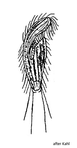
Kahl gives only a very short description and a drawing of Metopus setifer (s. drawing above). The specimens of my population showed a few deviations from his description. More than half of the specimens I examined were longer than 90 µm, but never reached 100 µm. I have not found any specimens smaller than 80 µm. The body shape of my specimens was also often oval and not tapered posteriorly (s. figs. 2 a-c and 3 a-c). The caudal cilia reached individually quite different lengths. In some specimens they measured about one third of the body length (s. fig. 1 a-c) while in others they were longer than the body (s. fig. 4 a-c). Very characteristic of Metopus setifer is the cilia-free terminal cell pole. The caudal cilia arise laterally around the posterior cell pole up to the posterior third, but not at the cell pole, which is thus naked (s. fig. 3 a). Kahl does not mention this feature in his description, but reproduces it in his drawing (s. drawing above).
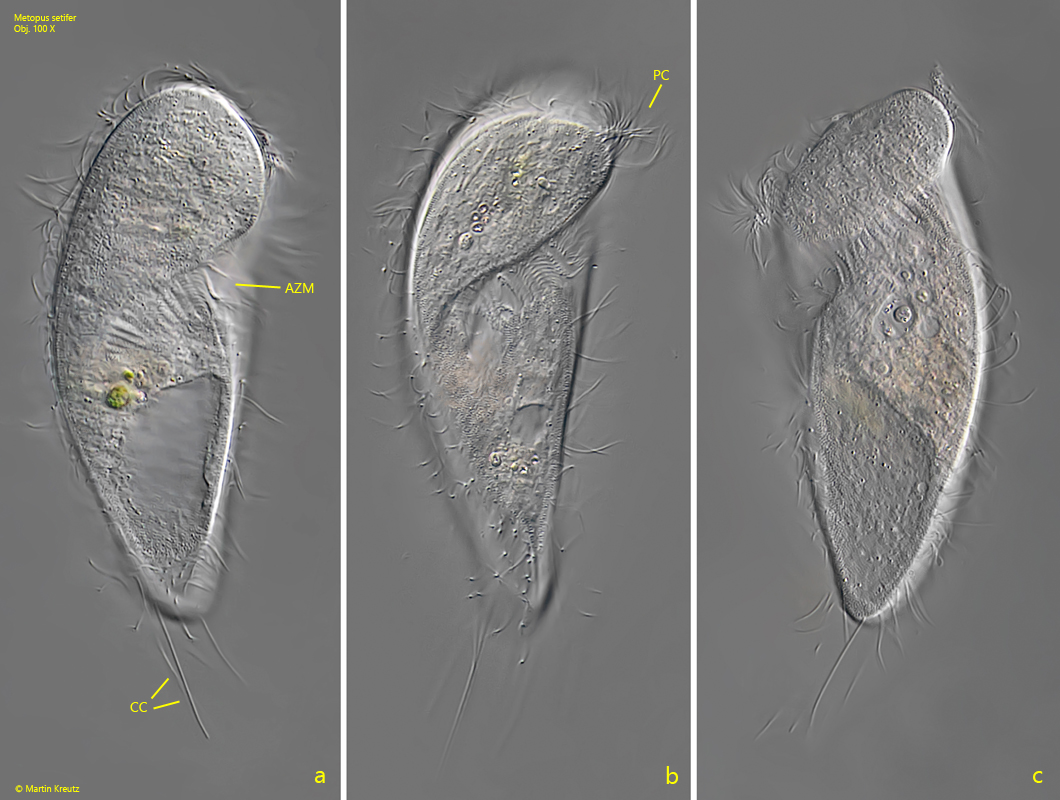
Fig. 1 a-c:Metopus setifer. L = 98 µm. A freely swimming specimen from ventral (a, b) and from the left side (s). AZM = adoral zone of membranelles, CC = caudal cilia, PC = perizonal cilia. Obj. 100 X.
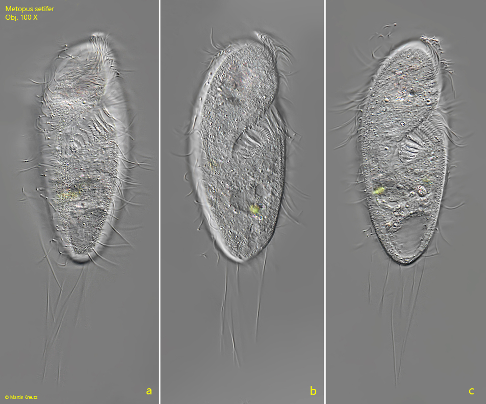
Fig. 2 a-c:Metopus setifer. L = 84 µm. A second, oval shaped specimen from ventral with longer caudal cilia. Obj. 100 X.
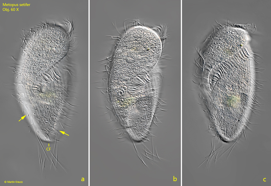
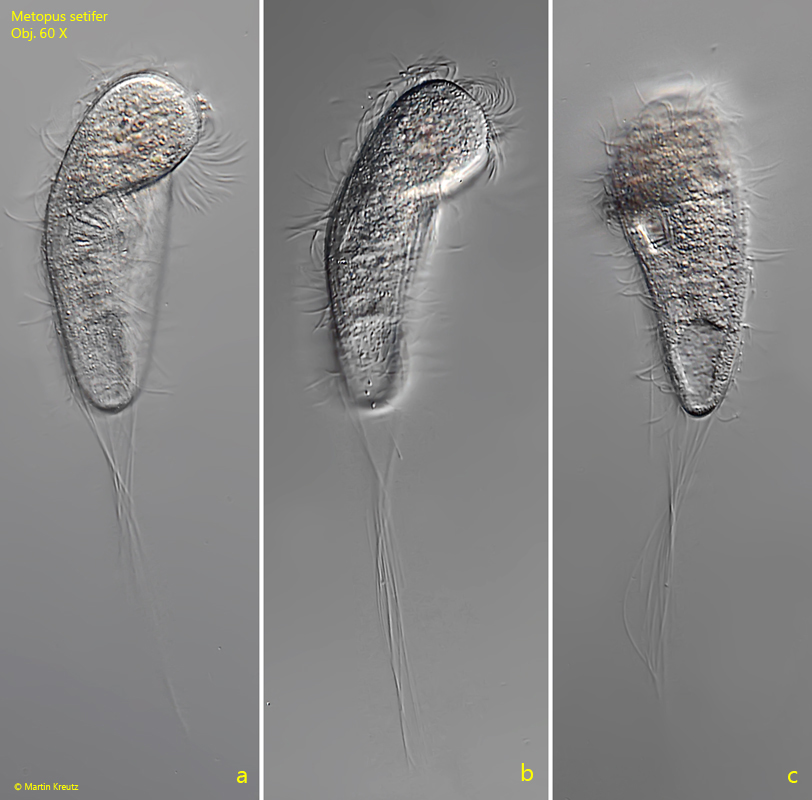
Fig. 3 a-c:Metopus setifer. L = 95 µm. A third, oval shaped specimen from ventral. Note that the caudal cilia arise at the posterior third (arrows) but not at the terminal pole. The posterior end is free from cilia (CF). Obj. 60 X.
Fig. 4 a-c:Metopus setifer. L = 84 µm. A freely swimming specimen with exceptionally long caudal cilia photographed at high layer thickness. The caudal cila are 87 µm long. Obj. 60 X.