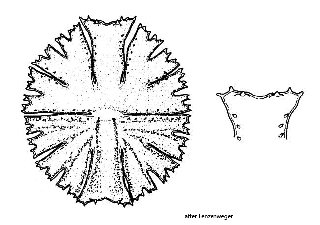
along the incisions of the lobes rows of short spines
Micrasterias papillifera
I found Micrasterias papillifera in June 1995 in Jackl Moor (Austria), in June 1998 in Rotmoor (Austria), in September 2007 in Walchsee (Austria) and most recently in June 2024 in the Paradieswiesen (Austria). I have not yet been able to find Micrasterias papillifera in the vicinity of Lake Constance. The specimen shown in fig. 1 a-b was sampled from the Walchsee (Austria) and the other shown specimens are from the Paradieswiesen.
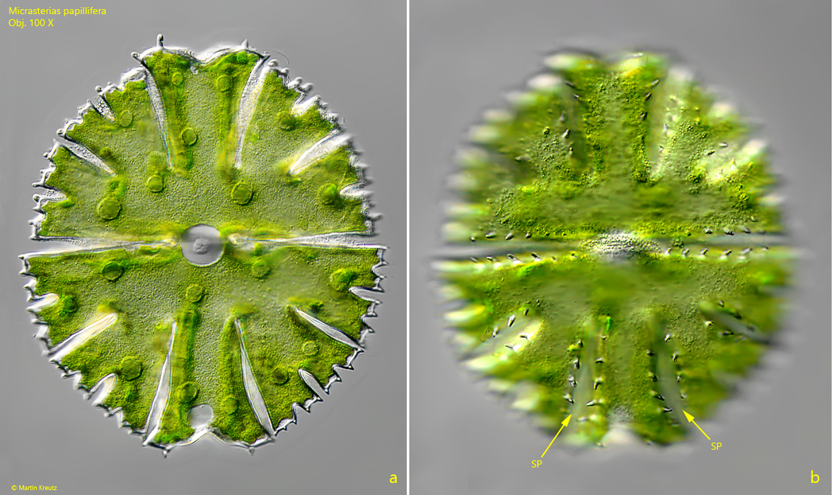
Micrasterias papillifera looks similar to Micrasterias rotata at low magnifications, but Micrasterias papillifera is only half the size. The main characteristic of Micrasterias papillifera are small spines, which are arranged in rows along the incisions between the lobes (s. figs. 1 b and 2 b).
Fig. 1 a-b:Micrasterias papillifera. L = 127 µm. Two focal planes of a specimen from Walchsee (Austria). Note the rows of short spines (SP) along the incisions of the lobes. Obj. 100 X.
Fig. 2 a-b:Micrasterias papillifera. L = 136 µm. Two focal planes of a specimen from the Paradieswiesen. SP = spines along the incisions of the lobes. Obj. 60 X.
Fig. 3 a-b:Micrasterias papillifera. L = 136 µm. The specimen as shown in fig. 2 a-b in detail. Obj. 100 X.
Fig. 4 a-b:Micrasterias papillifera. L = 121 µm. A second specimen from the Paradieswiesen. Obj. 100 X.