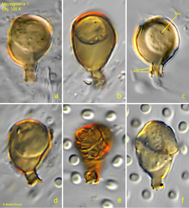
shell hyaline, colored orange-brown, without iron precipitation on older specimens
long neck, obliquely oriented to shell outline, annulated, distal end with thickened rim
nucleus central
contractile vacuole near neck
No drawings from previous authors available.
Microgromia 1 I have found exclusively in the Simmelried and this only until 2007. After that I have found no more specimens. This species is clearly different from the Microgromia species described by de Saedeleer. The shell of this species is always orange-brown colored, but without obvious iron deposits as in Microgromia haeckeliana. The neck is clearly angled. Its length is about a quarter of the shell length. It is also distinctly annulated, giving it a wavy appearance in optical section. The cell is often placed at the dorsal end of the shell and a long cytoplasm peduncle leads to the neck. I could not definitively detect a septum, as I only had a dry condenser in 2007, which prevented me from achieving the necessary resolution for detection. However, the described characteristics suggest this species to be a representative of the genus Microgromia.
Fig. 1:Microgromia 1. L = 14–20 µm. Six different specimens. All of them are colored orange-brown. Note the annulated neck (AN) of this species. Nu = nucleus. Obj. 100 X.
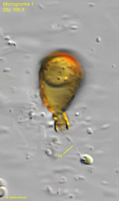
Fig. 2:Microgromia 1. L = 17 µm. This specimen tries to ingest a small alga caught with a thin granuloreticulopodium. Obj. 100 X.
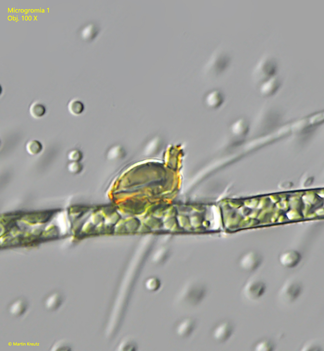
Fig. 3:Microgromia 1. L = 14 µm. A specimen attached to an alga filament. Note the almost vertically angled neck of the sessile specimen. Obj. 100 X.