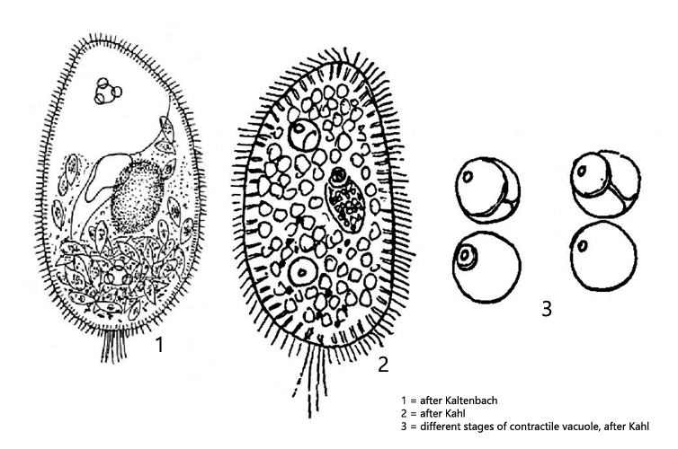
macronucleus ellipsoid with an attached large micronucleus
oral opening near mid-body
two contractile vacuoles with confluent auxiliary vacuoles
pellicle square fielded
fringe of spindle shapes extrusomes
no symbiotic algae
often light-refracting excretory crystals in posterior third
tuft of caudal cilia
Paramecium putrinum
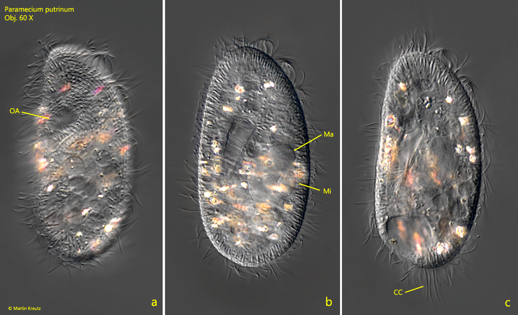
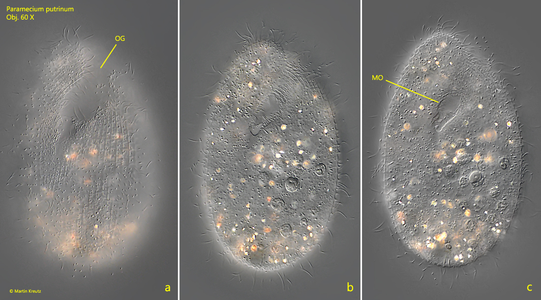
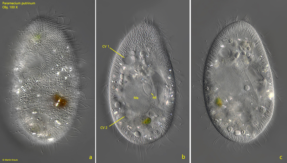
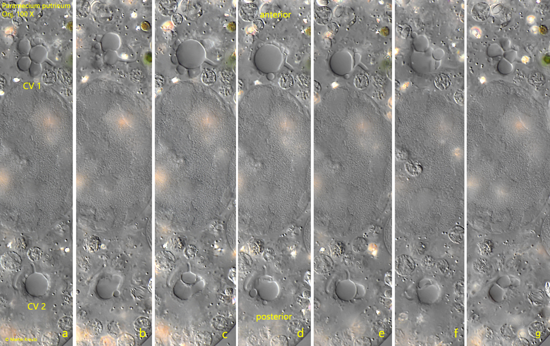
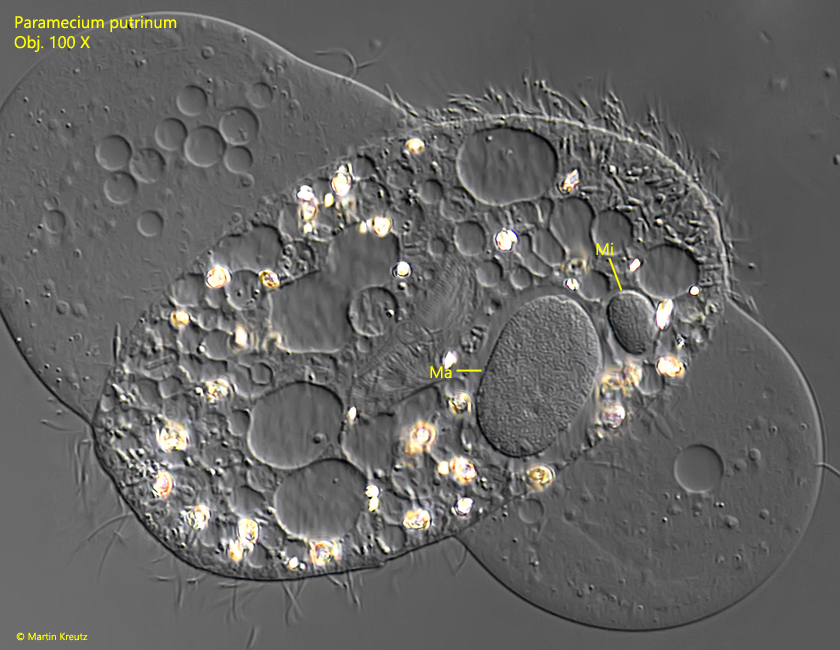
I find Paramecium putrinum sporadically between decaying plant masses from the Simmelried. In nutrient-rich and heavily polluted waters Paramecium putrinum should be much more common. Paramecium putrinum has a mouth opening typical of the genus Paramecium. A shallow groove leads to the mouth opening (s. fig. 2 a-c). Compared to Paramecium caudatum, the species is much smaller (mostly 80 – 100 µm) and the body is more stocky. Highly refractive crystals are frequently found in the plasma, which light up strongly in DIC and often aggregate in the posterior third of the body. However, no symbiotic algae are present, which distinguishes Paramecium putrinum from Paramecium bursaria. Two contractile vacuoles are present, which are often difficult to see in freely swimming specimens because they are located in deeper plasma layers connected to the excretory pores via tubes. The contractile vacuoles are surrounded by small auxiliary vacuoles that fuse even before the contractile vacuole has emptied. This can give the impression that two contractile vacuoles are adjacent to each other (s. fig. 4 a-g). At the posterior end, there is a tuft of caudal cilia about 10–15 µm long. A rather large micronucleus is closely attached to the ellipsoid macronucleus (s. fig. 5).
Fig. 1 a-c:Paramecium putrinum. L = 102 µm. Three focal planes of a freely swimming specimen from ventral. CC = tuft of caudal cilia, Ma = macronucleus, Mi = micronucleus, OA = oral apparatus. Obj. 60 X.
Fig. 2 a-c:Paramecium putrinum. L = 110 µm. Three focal planes from ventral of a slightly squashed specimen. Note the oral groove (OG) leading to the mouth opening (MO). Obj. 60 X.
Fig. 3 a-c:Paramecium putrinum. L = 83 µm. Ventral view (a) and dorsal view (b, c) of a freely swimming specimen. The two contractile vacuoles (CV 1, CV 2) a hard to see in a freely swimming specimen, because they are located deep in the cytoplasm and connected via tubes with the excretion pores. Obj. 100 X.
Fig. 4 a-g:Paramecium putrinum. Different stages of the two contractile vacuoles CV 1 and CV 2 photographed within 24 sec in a squashed specimen. Obj. 100 X.
Fig. 5:Paramecium putrinum. The macronucleus (Ma) und micronucleus (Mi) in a strongly squashed specimen. Obj. 100 X.