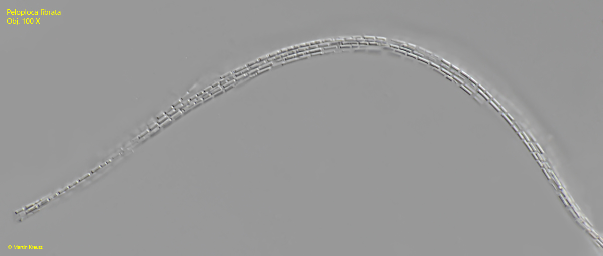
aggregates of cells are multilayered bundles of filaments up to 1 mm long
ends of the aggreates irregularly tapered or defibered
single bacteria cells are arranged tightly in the long filaments
each filament with an indistinct hyaline sheath
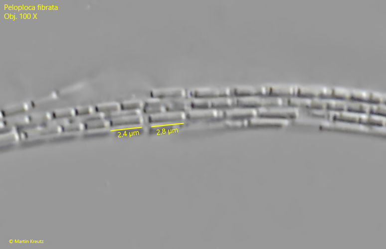
boundaries of the cylindrical single cells in the filaments indistinct, sometimes not visible
length of cells variable, up to 5 time longer than wide
cells hyaline, without gas vacuoles
Peloploca fibrata
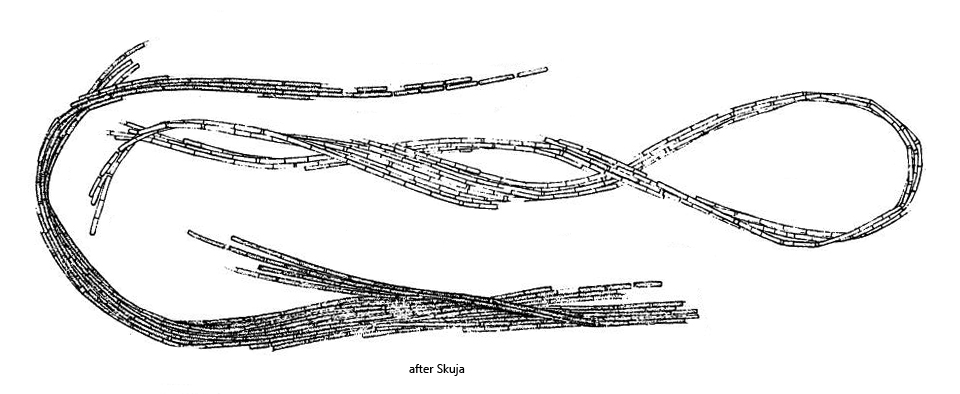
Peloploca fibrata is an aggregate-forming bacterium that was first described in 1956 by Skuja in Swedish Lapland. In the aggregates, which can consist of more than 1000 individual cells, the bacteria are arranged in filaments. These filaments in turn are arranged in bundles of varying thickness and length to form the aggregate (s. figs. 1 a-b, 2 and 3).
I find Peloploca fibrata regularly, but never frequently in the Simmelried. I have not yet been able to find this bacterium in my other sampling sites. Peloploca fibrata is almost always associated with rhodobacteria. Since the aggregates of Peloploca fibrata are very long (up to 1 mm), they are immediately noticeable in the samples even at low magnifications. In the aggregates of my population, the individual cells in the filaments were not or only indistinctly recognizable, as Skuja has already described. I only found a few aggregates in which the boundaries of individual cells could be clearly seen (s. figs. 4 and 5). They were almost always aggregates with only a few filaments. The individual cells may be easier to recognize in the growth phase of the aggregates. In such aggregates it can be seen that the cylindrical bacteria in the filaments have very different lengths, which is also described by Skuja. Important characteristics of Peloploca fibrata are that there are no constrictions between the individual cells and that the cells do not contain gas vacuoles. This distinguishes Peloploca fibrata from Peloploca taeniata.
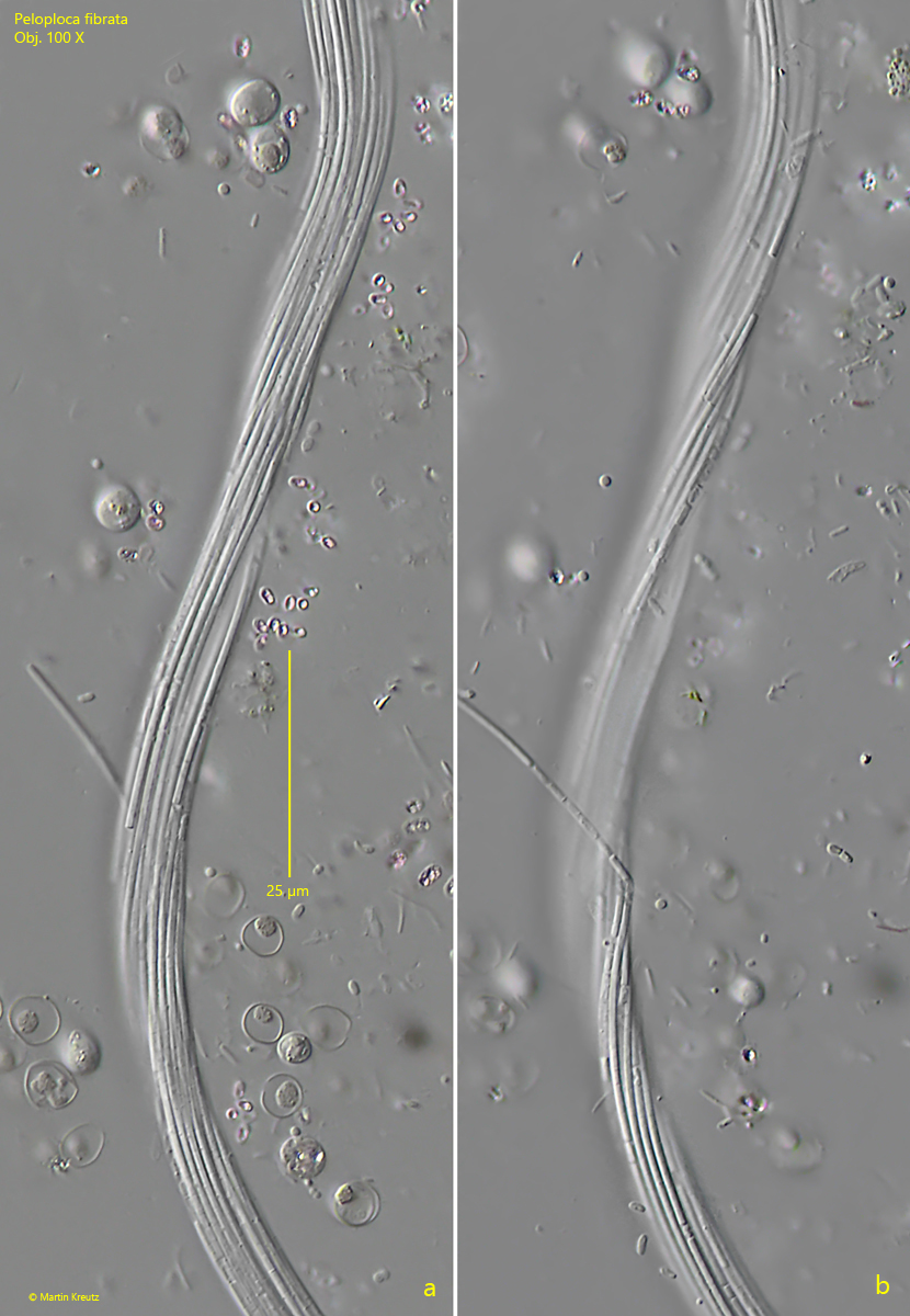
Fig. 1 a-b:Peloploca fibrata. L = about 500 µm (of the aggregate). Two focal planes of a slightly twistes aggreate of about 20–30 filaments. The boundaries of the single bacteria cells are only visible in few parts of the aggregate. Obj. 100 X.
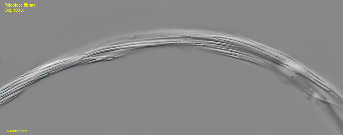
Fig. 2:Peloploca fibrata. L = about 600 µm (of the aggregate). A section of a second aggregate. Obj. 100 X.
Fig. 3:Peloploca fibrata. L = about 500 µm (of the aggregate). A section of a third aggregate. Obj. 100 X.
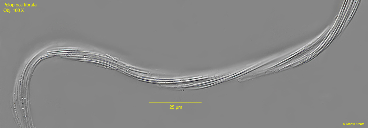
Fig. 4:Peloploca fibrata. L = about 300 µm (of the aggregate). In this aggregate of about 10 filaments the single bacteria cells in the filaments were clearly separated. Probably the separation of the cells is more easily visible at the beginning of aggregate formation. Obj. 100 X.
Fig. 5:Peloploca fibrata. In this close-up of the fig. 4 the different lengths of the bacteria in the filaments becomes visible. Note that the cells are hyaline without gas vacuoles. Obj. 100 X.