aggregates of cells are multilayered bundles of filaments up to 1 mm long
aggreates end in single filaments
single bacteria cells are arranged tightly in the long filaments
filaments without hyaline sheath, aggregates with 5 µm thick hyaline sheath
cross walls between cells slightly constricted, hard to see
single cells 0.7–1 µm x 3–10 µm with distinct gas vacuoles
Peloploca taeniata
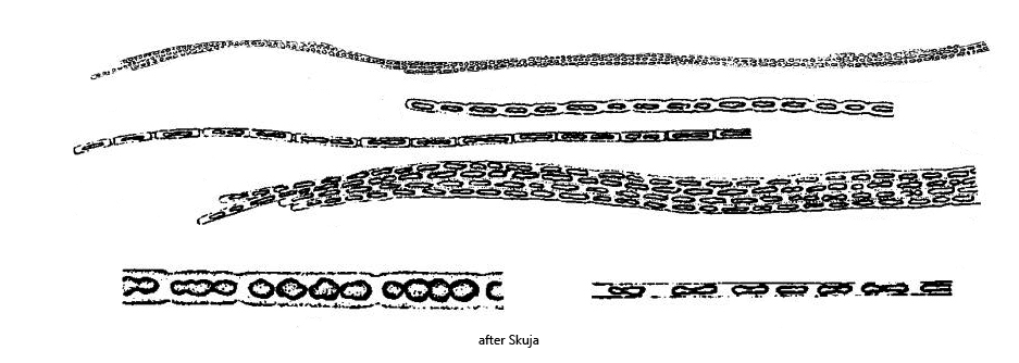
Peloploca taeniata is an aggregate-forming bacterium that was first described by Lauterborn in 1913. However, his description is very brief and not very informative. His drawings were also rather superficial. It was not until 1956 that Skuja published a re-description of this species in his results regarding the investigation of Swedish inland water bodies. His description is much more precise and his drawings more informative.
The aggregates of Peloploca taeniata consist of filaments of bacteria. These filaments in turn are arranged in bundles of varying thickness and length. The aggregates can therefore contain well over 1000 cells.
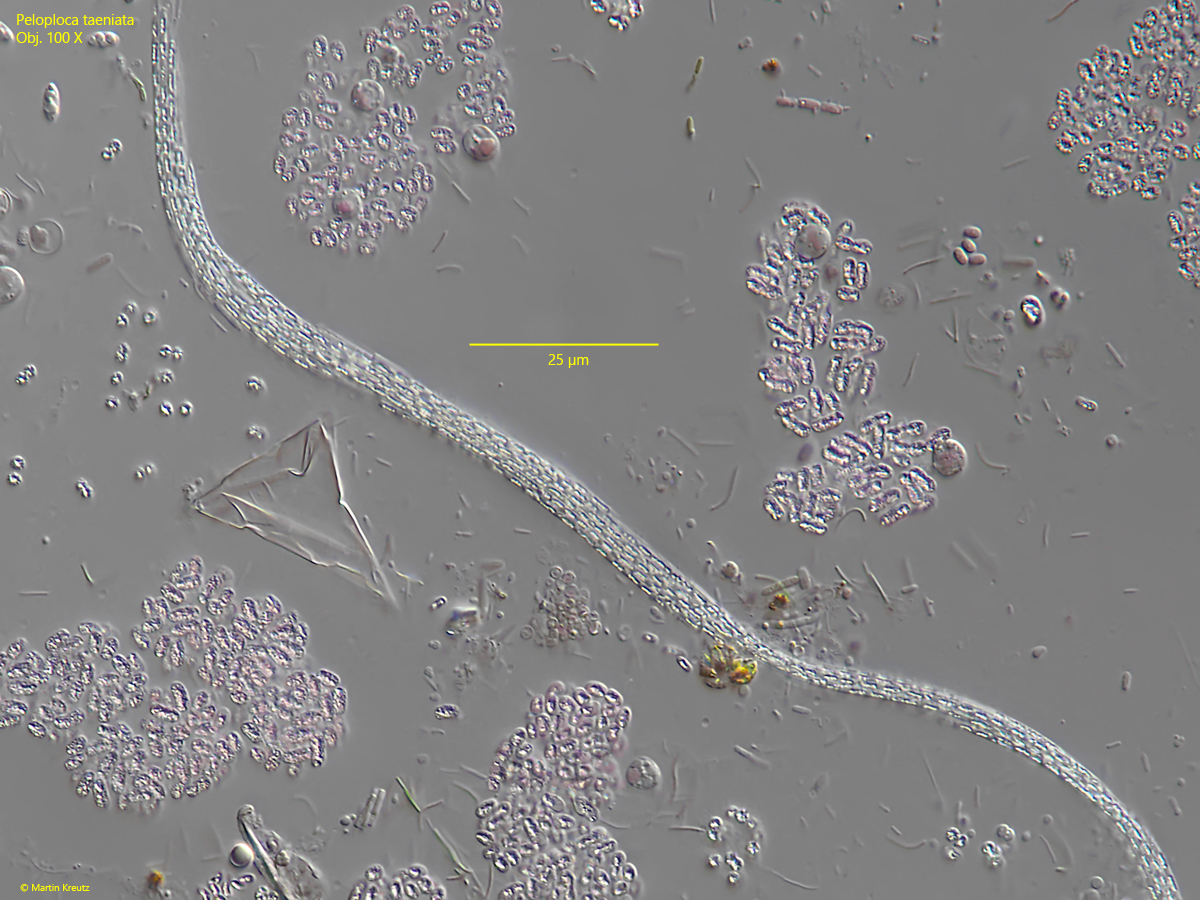
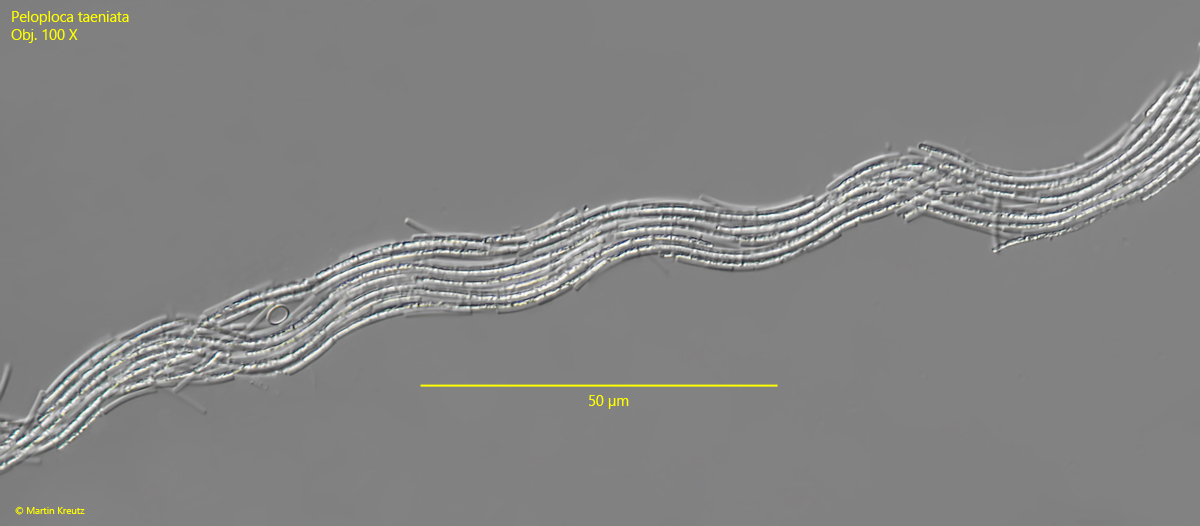
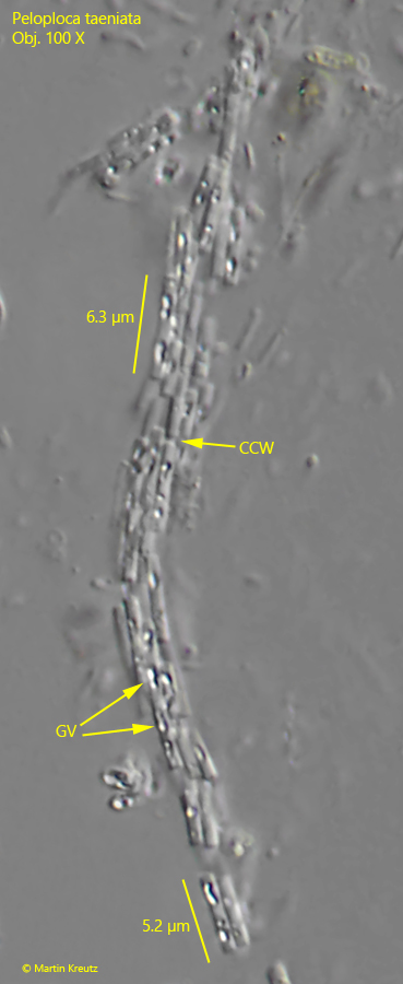
I find Peloploca taeniata regularly, but never frequently in the Simmelried. I have not yet been able to find this bacterium in my other localities. Peloploca taeniata is almost always associated with rhodobacteria (s. fig. 1). Since the aggregates of Peloploca taeniata are very long (up to 1 mm), they are immediately noticeable in the specimens even at low magnifications. The individual cells in the filaments were only clearly distinguishable in strongly squashed aggregates (s. fig. 4). According to my measurements, the individual cells are between 5–7 µm long, which fits well with the measurements of Skuja, who states 3–10 µm. Each cell contains one or two gas vacuoles, which gives the aggregates an almost glittering appearance. I often find the similar species Peloploca fibrata co-occuring with Peloploca taeniata in the samples. However, it is easy to distinguish the two species because Peloploca fibrata has no gas vacuoles and the cells are clear and hyaline.
Fig. 1:Peloploca taeniata. L = about 600 µm (of aggregate). An aggregate, consisting of a bundle of filaments, between other bacteria and rhodobacteria. Obj. 100 X.
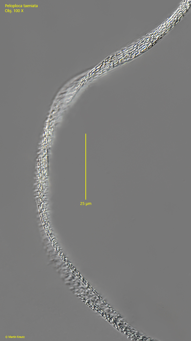
Fig. 2:Peloploca taeniata. L = about 700 µm (of aggregate). A slightly twisted aggregate. Due to the gas vacuoles in the cells the aggregate appears “glittering”. Obj. 100 X.
Fig. 3:Peloploca taeniata. L = about 500 µm (of aggregate). A more wavy and ribbon-shaped aggregate. Obj. 100 X.
Fig. 4:Peloploca taeniata. A severed piece of a squashed aggregate. Note the gas vacuoles (GV) in the cells and the slight constrictions of the cross walls (CCW) between the cells. Obj. 100 X.