contracted cells almost spherical, diameter 38–42 µm
stalk 140–320 µm long, contracts in spirals
cytoplasm colorless
two contracile vacuoles
macronucleus J-shaped
pellicle covered with pellicular tubercles.
vesicular tubercles closely-spaced, regularly arranged in 24–28 transverse rows
solitary or in pseudocolonies
No drawings from previous authors available.
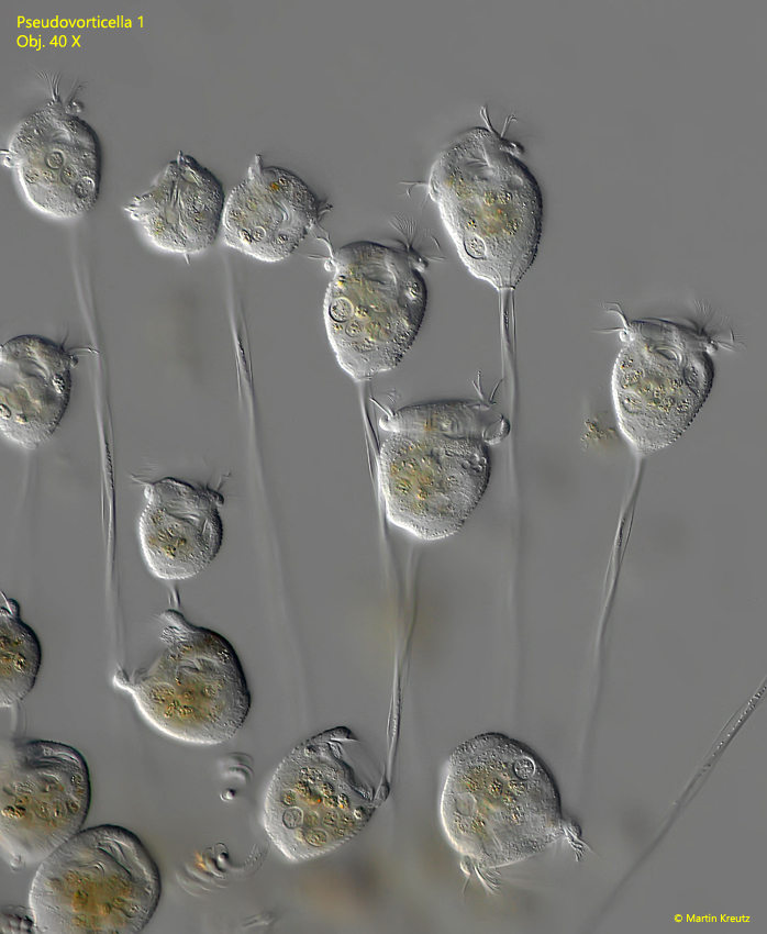
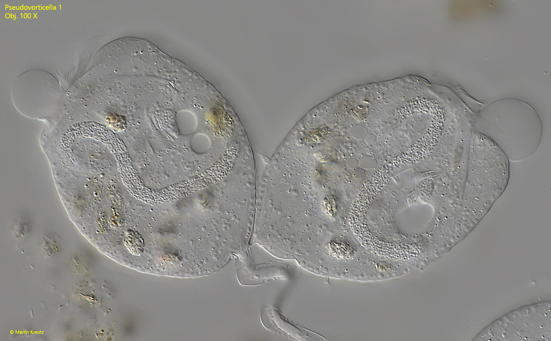
In August 2022 I found several pseudocolonies of a Pseudovorticella on floating detritus flakes in an old sample from the Simmelried. The individuals were 42–80 µm long, on 140–320 µm long stalks, which contracted in spirals. The contracted individuals were approximately spherical with a diameter of 38–42 µm. Already at medium magnification it was obvious that the pellicle was covered with regularly arranged vesicles.
Fig. 1:Pseudovorticella 1. L = 42–72 µm. Freely moving specimens of a pseudocolony. Obj. 40 X.
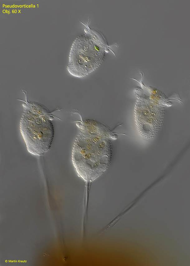
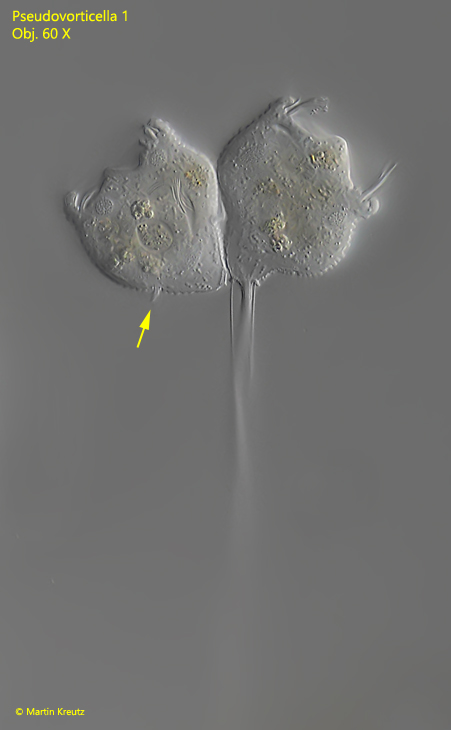
Fig. 2:Pseudovorticella 1. L = 56–62 µm. Freely moving specimens of a pseudocolony. Obj. 60 X.
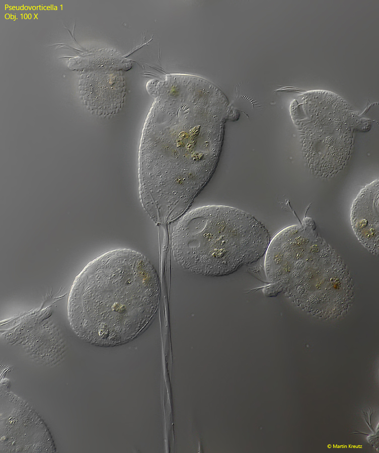
Fig. 3:Pseudovorticella 1. L = 80 µm. Freely moving specimens of a pseudocolony. Obj. 60 X.
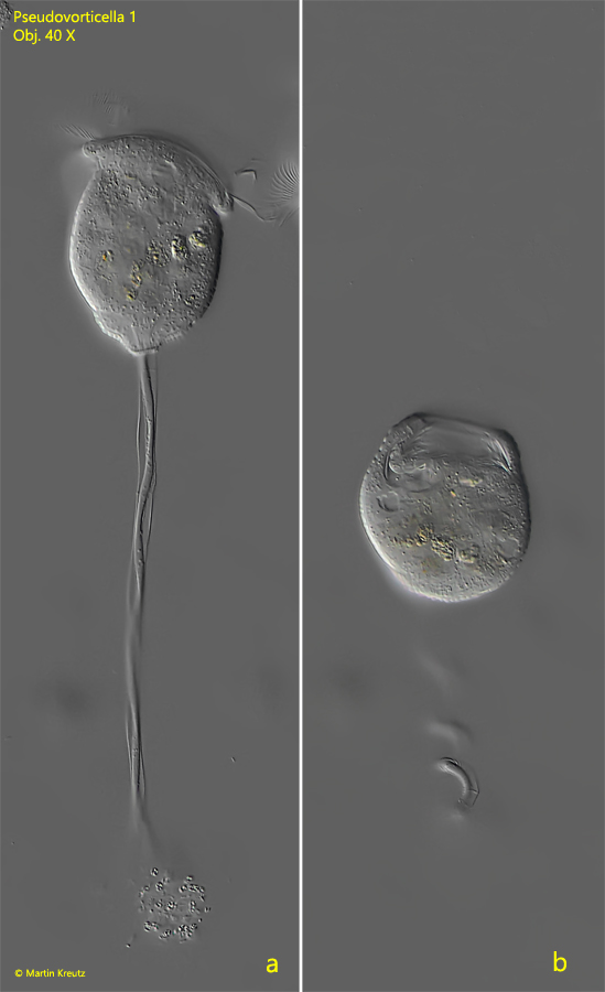
Fig. 4 a-b:Pseudovorticella 1. L = 67 µm. An extended specimen on a 165 µm long stalk (a) and the same specimen almost spherically contracted with a diameter of 40 µm (b). Obj. 40 X.
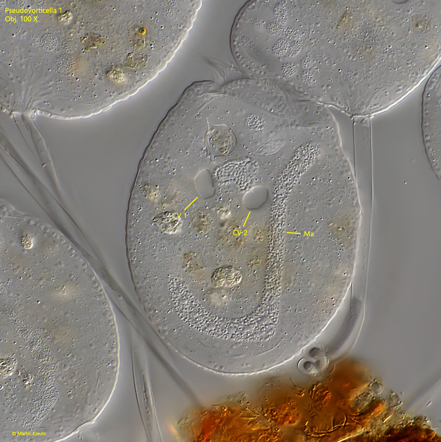
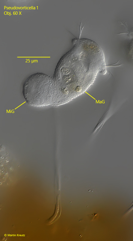
In a squashed specimen, it could be seen that the macronucleus is J-shaped and that two contractile vacuoles are present.
Fig. 5:Pseudovorticella 1. A squashed specimen with a J-shaped macronucleus (Ma) and two contractile vacuoles (CV 1, CV 2). Obj. 100 X.
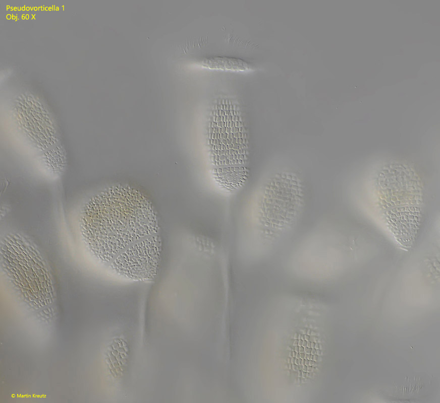
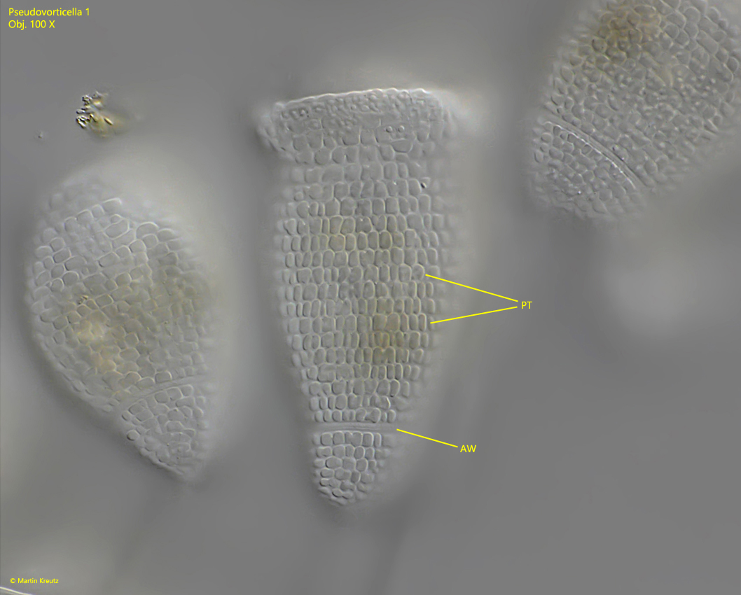
The pellicle of the individuals is covered with regularly arranged vesicles (pellicular tubercles). I could count 24–28 transverse rows of them, but the vesicles on the peristome were difficult to be recognized individually. The vesicles are so densely arranged that they touch each other. They deform at the contact surfaces, which can sometimes result in polygonal vesicles. Essentially, however, the vesicles are broadly oval and with their long axis oriented in parallel to the long axis of the cell. Some vesicles appeared to be in a state of division and had indentations at their apices. The content of the vesicles was always clear and colorless.
Fig. 6:Pseudovorticella 1. Focus on the pellicular tubercles covering the pellicle of freely moving specimens in a pseudocolony. Obj. 60 X.
Fig. 7:Pseudovorticella 1. Detail of the pellicular tubercles (PT) in a slightly squashed specimen. AW = aboral ciliary wreath. Obj. 100 X.
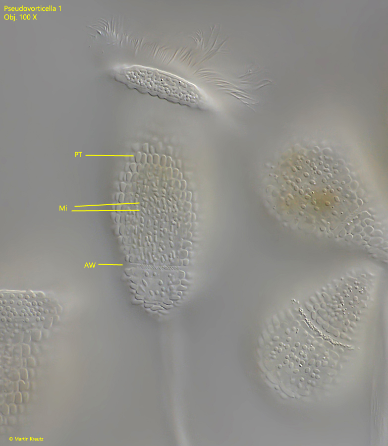
Fig. 8:Pseudovorticella 1. The same specimen as in fig. 7 with a slightly lowered focal plane. The mitochondria (Mi) arranged beneath the pellicle becomes visible. AW = aboral ciliary wreath, PT = pellicular tubercles. Obj. 100 X.
Given the shape and arrangement of the well defined pellicular tubercles (blisters) this species cannot be Pseudovorticella monilata with globular tubercles, often filled with refractive oily droplets. There only few alternatives of described Pseudovorticella species with two contractile vacuoles. The species Pseudovorticella sphagni and Pseudovorticella mollis can be excluded, because they are much smaller (max. length 50 µm) and the shape of the body is different. There are no further alternatives. Therefore I’m convinced that this represents an undescribed species (s. most likely ID = Pseudovorticella nov. spec.).
Within the pseudocolonies of Pseudovorticella 1 I could observe many stages of cell division. The daughter cell is growing an aboral ciliary ring, becomes cylindrical and swims away as a so called telotroch.
Fig. 9:Pseudovorticella 1. A specimen in the process of binary cell fission. The daughter cell (left) is developing a ciliary ring (arrow) in the region of the aboral ciliary wreath. Obj. 100 X.
Fig. 10:Pseudovorticella 1. A squashed pair of cells in the process of binary fission. The daughter cell (right) is connected with the parent cell with a thin ctoplasmic bridge near the origin of the stalk. Obj. 100 X.
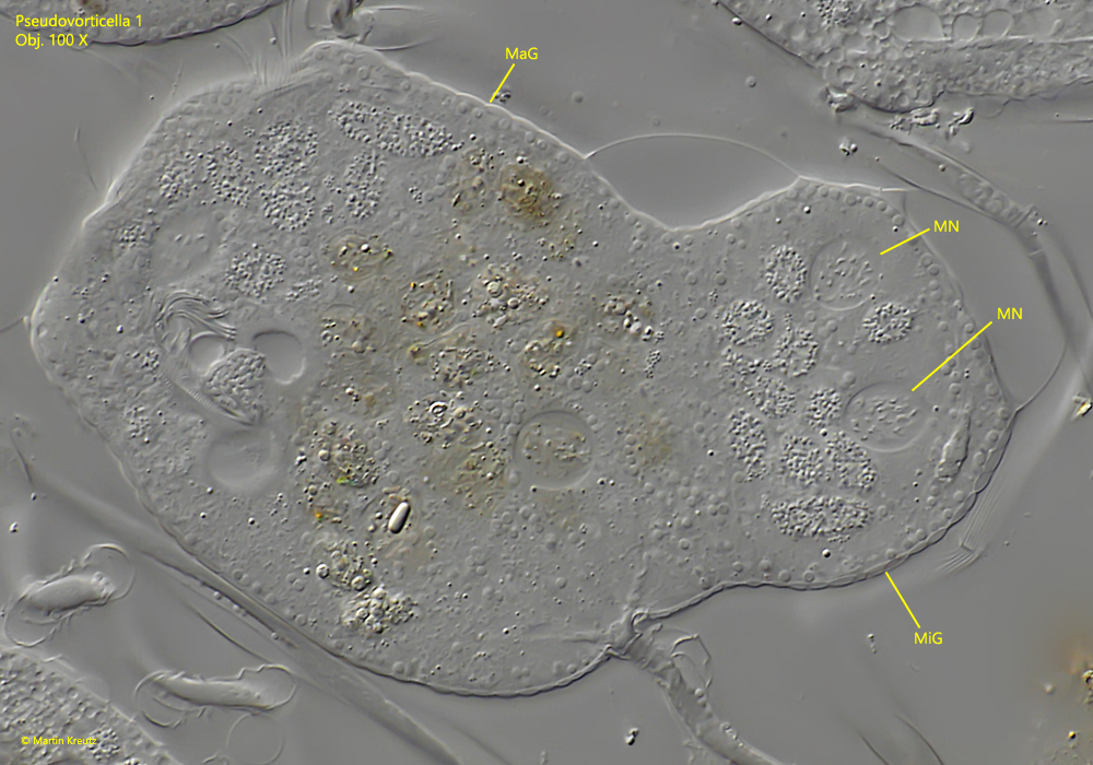
Besides cell divisions, stages of sexual reproduction were also found in the pseudocolonies (conjugaction). This takes place in sessile peritrichs by the fusion of a mobile microgamete with the sessile macrogamete (the sessile zooid). It can be recognized microscopically by the fact that the microgamete is much smaller than the macrogamete and fuses with it. As a result, macrogametes are found giving the impression of having a strong bulge at the posterior end.
Fig. 11:Pseudovorticella 1. The process of conjugation between a microgamete (MiG) and the sessile macrogamete (MaG). The microgamete is almost completely fused with the macrogamete and visible as a laterally bulge. Obj. 60 X.
Once the microgamete is fused with the macrogamete near the origin of the stalk, the macronuclei in the both cells begin to disintegrate. At the same time, the two micronuclei in both cells begin a meiotic reduction division. The resulting haploid micronuclei later fuse in the macrogamete and then the re-creation of a new macronucleus starts. I could not follow the whole process of conjugation, but different stages were found in the pseudocolonies, where the meiotic reduction division as well as the degradation of the macronucleus were well visible (s. figs. 12 -16).
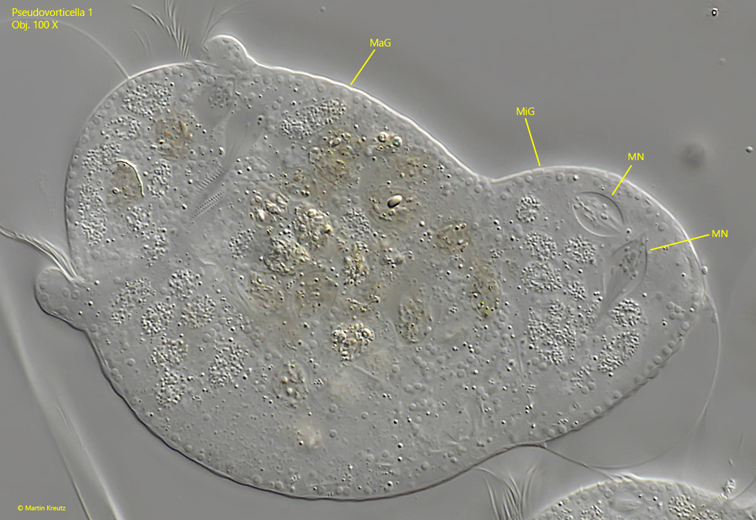
Fig. 12:Pseudovorticella 1. Process of meiotic division of micronuclei during conjugation of a microgamete (MiG) and the sessile macrogamete (MaG). In the meiotic micronuclei (MN) the condensed chromosomes are visible. Obj. 100 X.
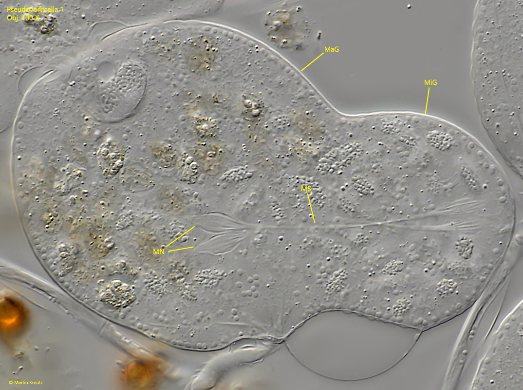
Fig. 13:Pseudovorticella 1. Process of meiotic division of micronuclei during conjugation of a microgamete (MiG) and the sessile macrogamete (MaG). The meiotic spindle apparatus (MS) in the meiotic micronuclei (MN) is visible to separate the chromosomes. Obj. 100 X.
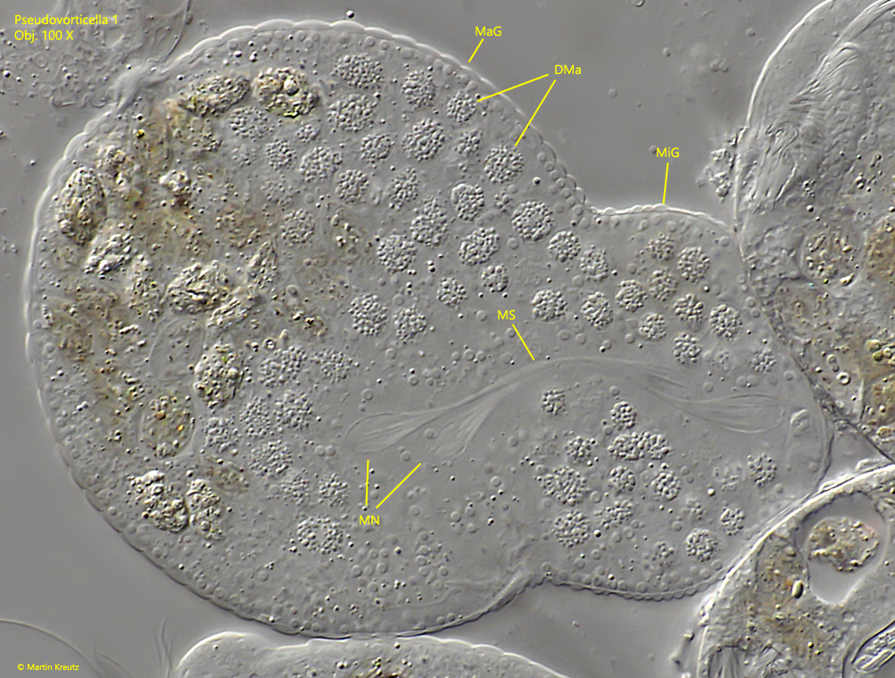
Fig. 14:Pseudovorticella 1. Process of meiotic division of micronuclei during conjugation of a microgamete (MiG) and the sessile macrogamete (MaG). The meiotic spindle apparatus (MS) in the meiotic micronuclei (MN) is visible to separate the chromosomes. Also the remains of the disintegrated macronucleus (DMa) are visible. Obj. 100 X.
Fig. 15:Pseudovorticella 1. Process of meiotic division of micronuclei during conjugation of a microgamete (MiG) and the sessile macrogamete (MaG). The meiotic spindle apparatus in the meiotic micronuclei (MN) is visible to separate the chromosomes. Obj. 100 X.
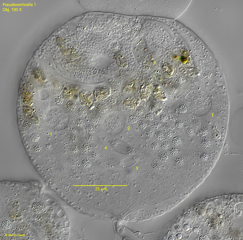
Fig. 16:Pseudovorticella 1. In this specimen the microgamete is completely fused with the macrogamete. The macronuclei of both cells are almost completely disintegrated. In the cell 5 micronuclei are visible in different stages of a meiotic division (1–5). In the micronuclei no. 4 and no. 5 the chromosomes are being separated by the meiotic spindle apparatus. Obj. 100 X.