cells about 20–40 µm in diameter (without coat of scales)
scales 7.2–12.7 µm x 3.2–4.8 µm, shaped oblong with parallel sides
edges of the scales strongly inflected
edges of the scales with a distinct striation (septa)
scales do not cover the base of the axopodia
cells sometimes with (symbiotic?) algae
centroplast in the center of the cell
nucleus in eccentric position
2–3 contractile vacuoles
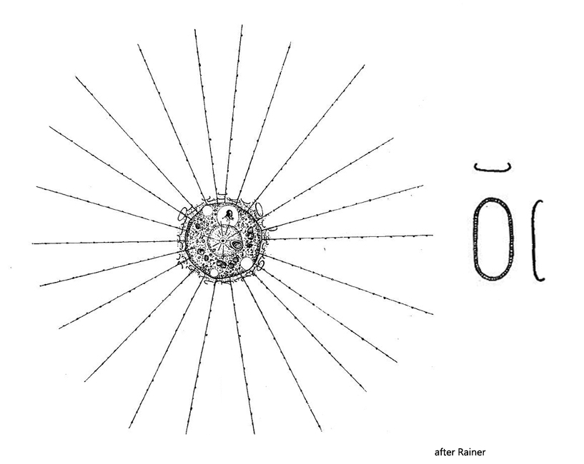
Raphidiophrys intermedia
I find Raphidiophrys intermedia quite often. The species lives solitarily and does not form colonies like Raphidiophrys elegans. The two species can be distinguished by the shape of the scales. In Raphidiophrys intermedia the scales are oblong (with parallel sides) and about twice as long as wide. They have a striped margin. In Raphidiophrys elegans, however, the scales are broadly oval.
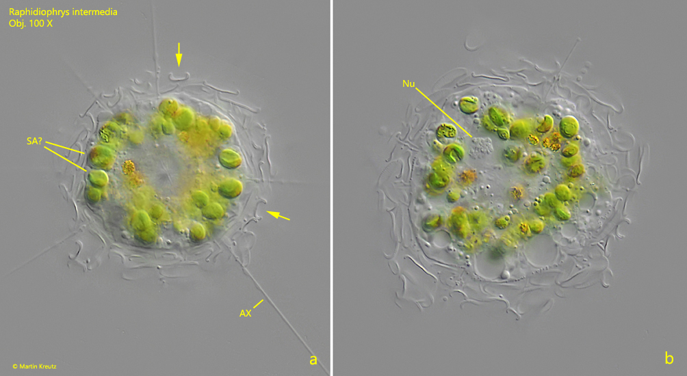
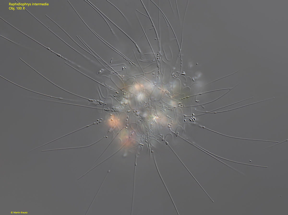
In my population of Raphidiophrys intermedia I often find specimens with algae in the cytoplasm (s. fig. 1 a-b). Rainer, 1968 (s. literature) mentions that the specimens may be sometimes green, but leaves open whether the green color is due to the presence of symbiotic algae. The algae in my specimens all appeared to be of the same species and placed in the periphery of the cell (s. fig. 1 a). They also did not appear to be inside food vacuoles and undergoing a digestive process. Therefore, I believe that the algae in the green specimens of Raphidiophrys intermedia are symbiotic algae.
Fig. 1 a-b:Raphidiophrys intermedia. D = 38 µm (without coat of scales). Two focal planes of a slightly squashed specimen with (symbiotic?) algae (SA?) in the cytoplasm. The scales have the shape of a “U” when optically sectioned along the longitudinal axis (arrows, a). AX = axodopdia, Nu = nucleus, SA = likely symbiotic algae. Obj. 100 X.
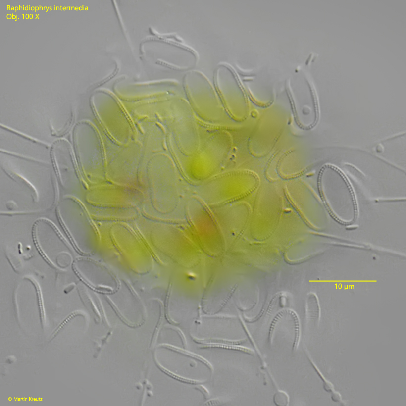
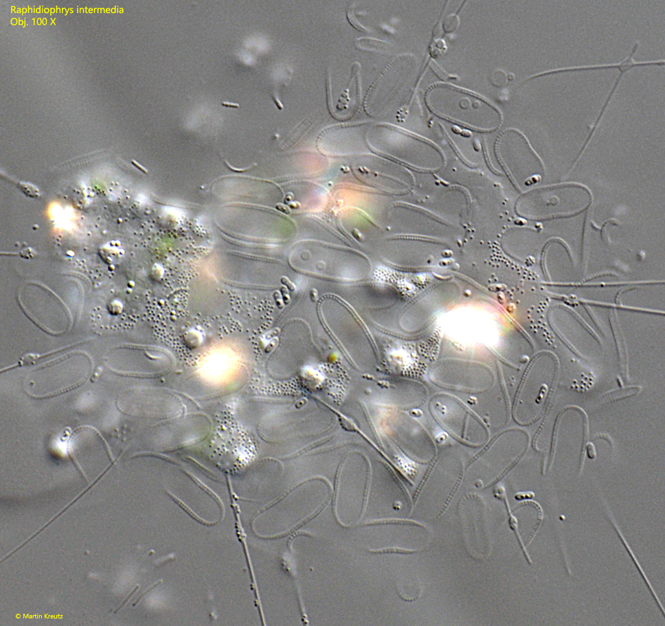
Fig. 2:Raphidiophrys intermedia. Focal plane on the coat of scales of the specimen shown in fig. 1 a-b. The oblong scales are about 10 µm long with a fine striation at the edges. Obj. 100 X.
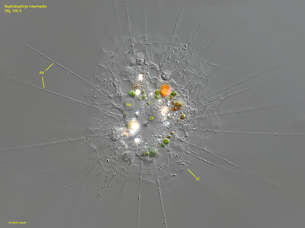
Fig. 3:Raphidiophrys intermedia. A strongly squashed specimen with focal plane on the centroplast (CP) in the center of the cell. AX = axopodia, Nu = nucleus, SC = scales. Obj. 100 X.
Fig. 4:Raphidiophrys intermedia. Focal plane on the coat of scales of the specimen shown in fig. 3. Obj. 100 X.
Fig. 5:Raphidiophrys intermedia. The scales of the specimen shown in figs. 3 and 4 in detail. Obj. 100 X.