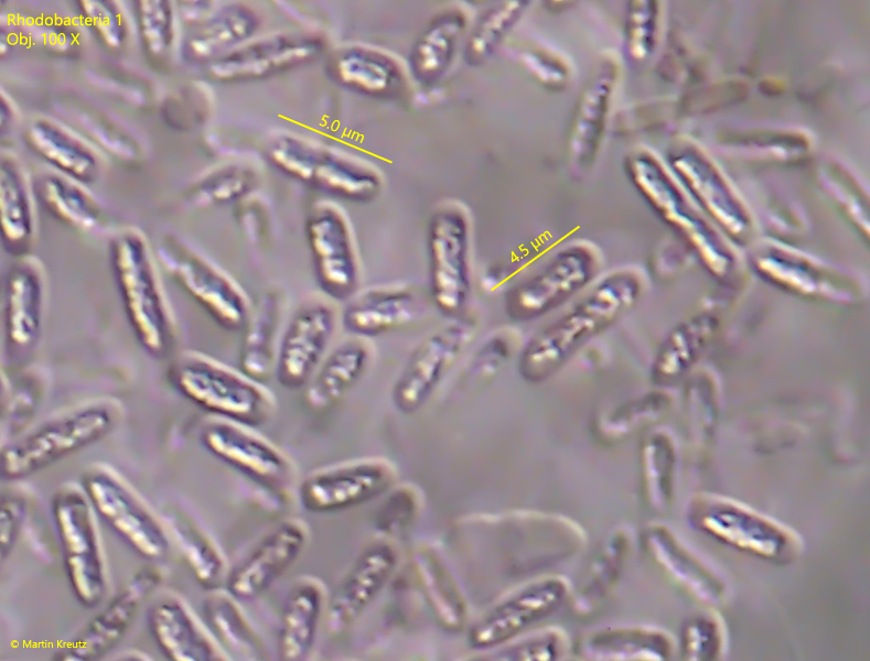
one irregularly shaped, highly refractive mass in center
color slightly pink
colonies irregularly shaped without sharp outline
no visible gelatinuous sheet
colonies of about 50–100 µm in diameter
No drawings from previous authors available.
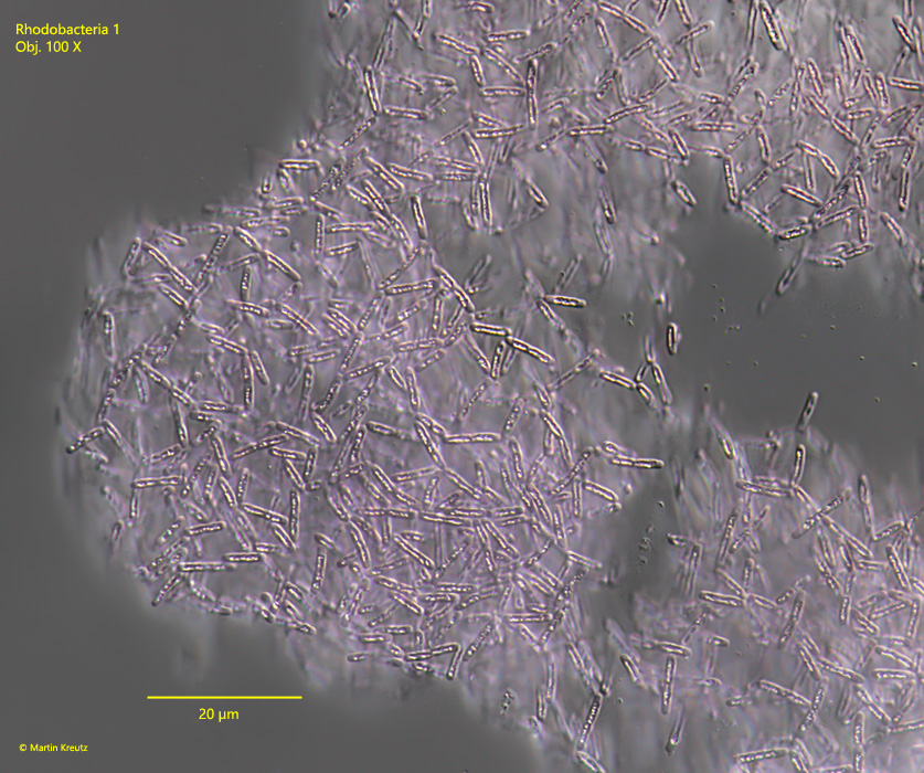
Fig. 1:Rhodobacteria 1. L = 4–5.5 µm. A not squashed colony. Obj. 100 X.
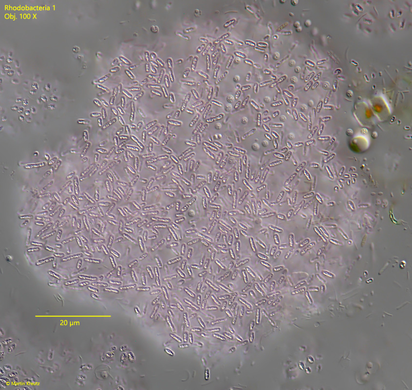
Fig. 2:Rhodobacteria 1. L = 4–5.5 µm. A pink colored colony with a diameter of about 80 µm. The individual cells are separated from each other and arranged irregularly in the colony. Obj. 100 X.
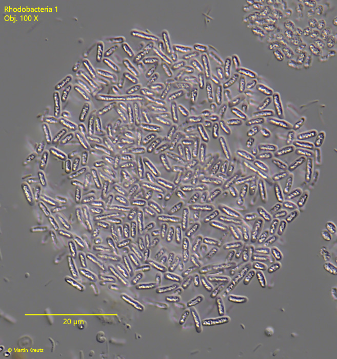
Fig. 3:Rhodobacteria 1. L = 4–5.5 µm. A slightly squashed, smaller colony. Obj. 100 X.
Fig. 4:Rhodobacteria 1. L = 4–5.5 µm. The single Rhodobacteria 1 cells in detail. Note the irregularly shaped, highly refractive mass in the center of each cell. Obj. 100 X.