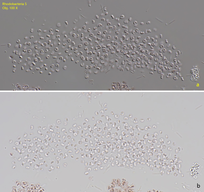
Fig. 1 a-b:Rhodobacteria 5. L = 2.6 X B = 2.3 µm. A slightly squashed colony in DIC (a) and brightfield illumination (b). All cells are separated from each other. Obj. 100 X.
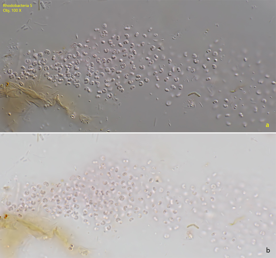
Fig. 2 a-b:Rhodobacteria 5. L = 2.6 X B = 2.3 µm. A second slightly squashed colony in DIC (a) and brightfield illumination (b). Obj. 100 X.
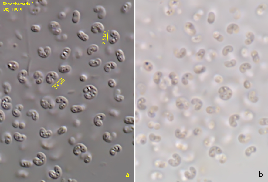
Fig. 3 a-b:Rhodobacteria 5. L = 2.6 X B = 2.3 µm. The U-shaped cells of Rhodobacteria 5 in detail in DIC (a) and brightfield illumination (b). Several spherical inclusions are always visible in the cells. Obj. 100 X.