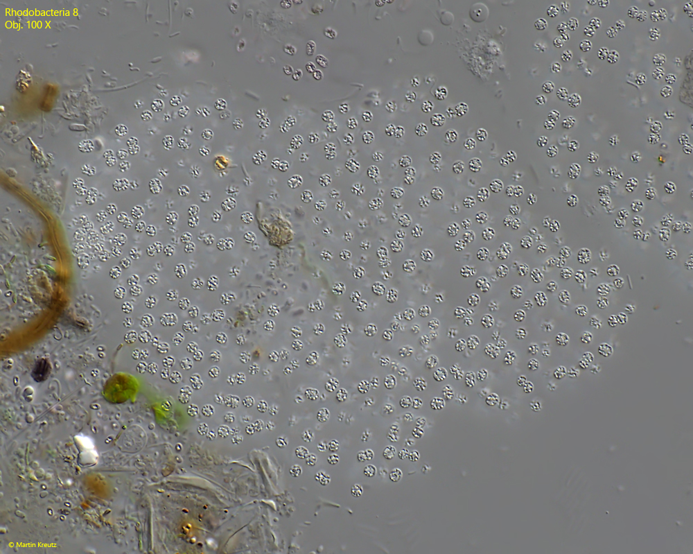
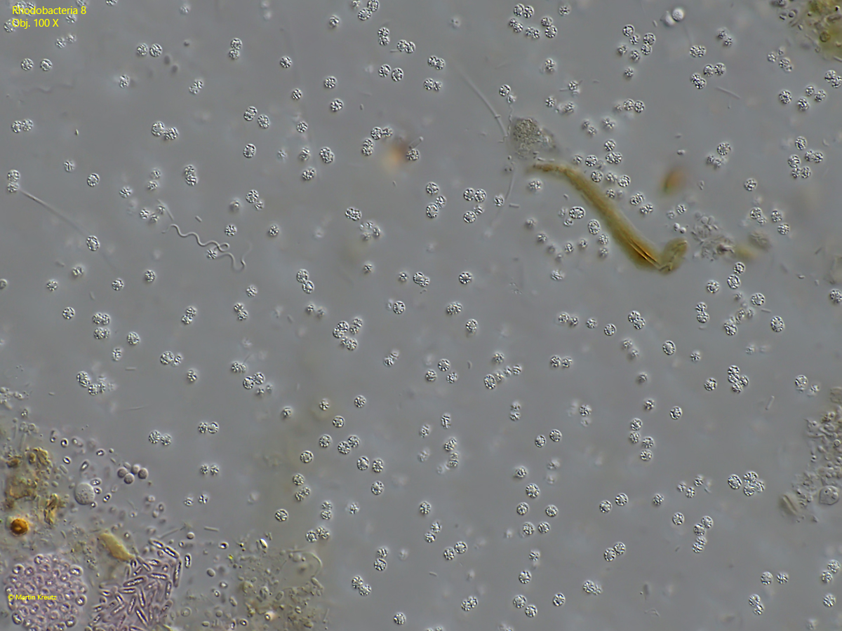
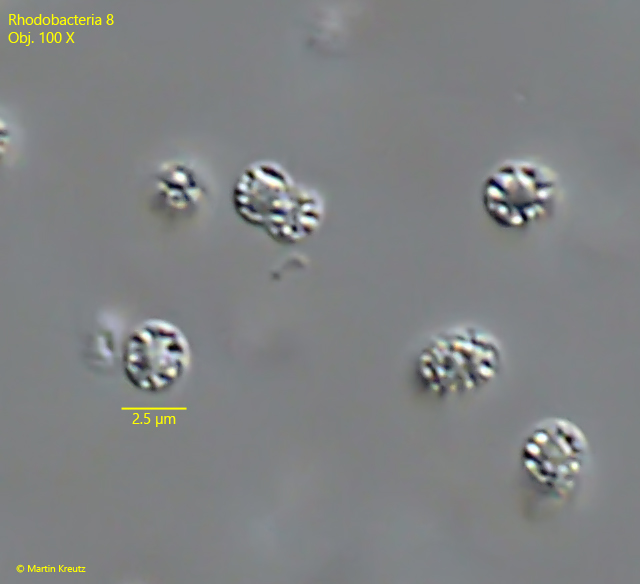
Rhodobacteria 8 Most likely ID: n.a. Synonym: n.a Sampling location: Simmelried Phylogenetic tree: n.a. Diagnosis:the cells are sphericaldiameter 2.2. – 2.6 µmhighly refractive mass mainly arranged in the periphery of cellscolorlesscolonies irregularly shaped without sharp outlineno visible gelatinous sheetcolonies of about 50 – 150 µm in diametercells in colony are sparated from each other No drawings from previous authors available. Fig. 1: Rhodobacteria 8. D = 2.2. – 2.6 µm. A slightly squashed colony. All cells are separated from each other. Obj. 100 X. Fig. 2: Rhodobacteria 8. D = 2.2. – 2.6 µm. A more strongly squashed colony. All cells are separated from each other. Obj. 100 X. Fig. 3: Rhodobacteria 8. D = 2.2. – 2.6 µm. The cells of a colony in detail. Note the refractive mass in the periphery of the cells. Obj. 100 X. Download PDF