right between ventral tooth and dorsal spine two spines
left two spines and the dorsal spine
frontal tooth short
one or two spherical macronuclei
contractile vacuole in posterior third
Saprodinium mimeticum var. simplex
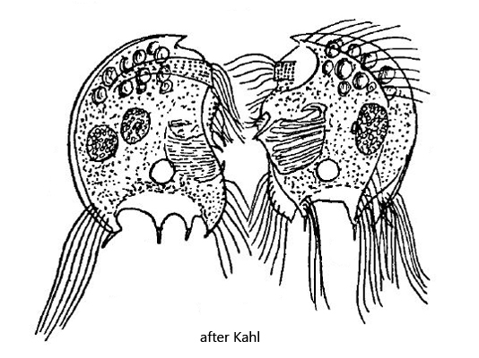
Kahl examined Saprodinium mimeticum very carefully and discovered variations with significant characteristics, which he described in the two subspecies Saprodinium mimeticum var. obliquum and Saprodinium mimeticum var. simplex. In my populations from the Simmelried I could also detect these form variantions described by him.
Saprodinium mimeticum var. simplex differs from the parent form by a much shorter frontal tooth. The ventral tooth is not long and curved but short and V-shaped. The dorsal spine is pointing dorsally and the median spines are shorter than in the parent form. Overall, the spines are shortened compared to the parent form. However, Saprodinium mimeticum var. simplex lacks the ventral tooth on the left side, as described for the parent form.
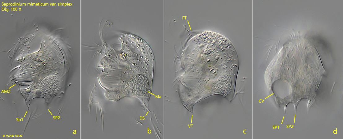
I could find the variant Saprodinium mimeticum var. simplex twice in Simmelried. Both times in November of the years 2020 and 2022. Unfortunately I could only take images from the left side. In the second specimen, however, I set the focal plane through the specimen on the right side (s. figs. 2c and 2d).
Fig. 1 a-c: Saprodinium mimeticum var. simplex. L = 38 µm. A freely swimming specimen from the left side. AZM = adoral zone of membranelles; DC = dorsal elongated cilia; DS = dorsal spine; Ma = macronucleus; SB = symbiotic bacteria; SP1, SP2 = median posterior spines left side. Obj. 100 X.
Fig. 2 a-d: Saprodinium mimeticum var. simplex. L = 42 µm. Four focal planes of a second specimen from the left side (a, b) to the right side (c, d). Note the short ventral tooth (VT) of the right side (s. fig. 2c). AZM = adoral zone of membranelles; DS = dorsal spine; CV = contractile vacuole; FT = frontal tooth; Ma = macronucleus; SP1, SP2 = median posterior spines left side; SP1′, SP2′ = median posterior spines right side, VT = ventral spine right side. Obj. 100 X.
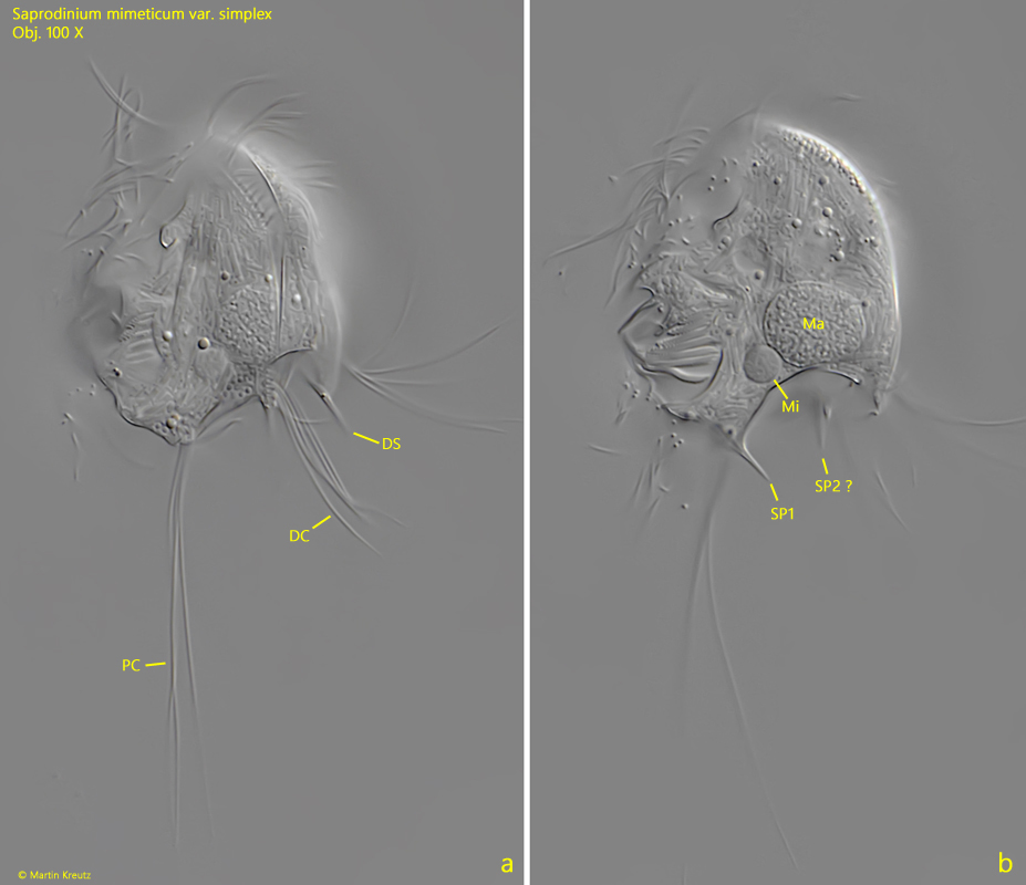
Fig. 3 a-d: Saprodinium mimeticum var. simplex. L = 51 µm. Note the tufts of long posterior (PC) and dorsal cilia (DC). The posterior cilia are 54 µm long. The curved ventral spine (VS) is visible and the dorsal spine (DS). AZM = adoral zone of membranelles, FT = frontal tooth, Ma = macronucleus, Mi = micronucleus. SP1 = median posterior spine. Obj. 100 X.