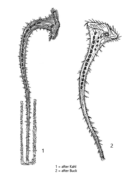
body elongated trumpet-shaped, contracted obovoid to club-shaped
appears yellowish or brownish, no symbiotic algae
length up to 3000 µm (of elongated specimens)
adoral membranelle running in clockwise direction to oral funnel
attached with thigmotactic cilia to the substrate
sometimes in a hyaline case
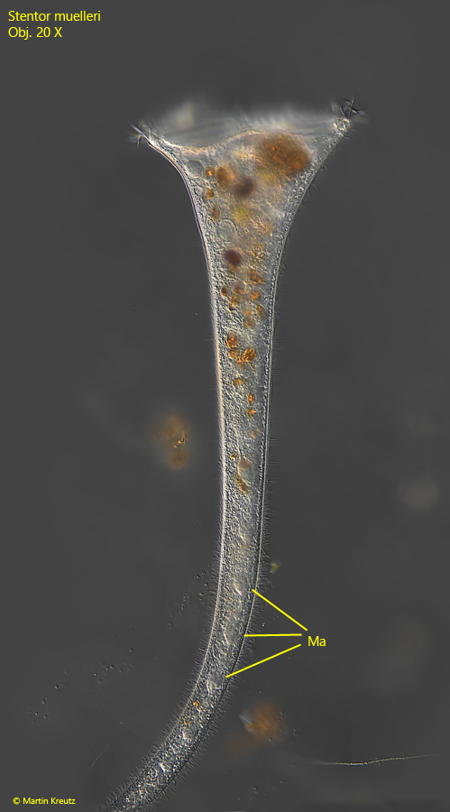
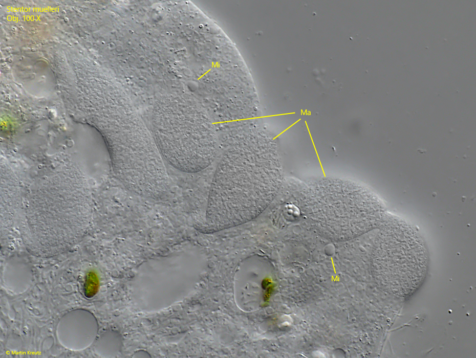
macronucleus moniliform with about 10–20 spherical parts
10–17 spherical micronuclei adjacent to the nodules of the macronucleus
contractile vacuole on left wall of oral funnel
Stentor muelleri
Stentor muelleri is one of the most common ciliates. I find it in practically all my sampling sites. It is easily recognizable by its mostly yellow-brown color and the moniliform macronucleus, which can be seen even in unsquashed specimens (s. fig. 4). The two other Stentor species with a moniliform macronucleus are Stentor coeruleus (colored blue or blue-green) and Stentor polymorphus (green due to symbiotic algae).
If a few specimens of Stentor muelleri are placed under a coverslip with a high layer thickness, they usually stretch out completely after a short time. The specimens in my population were between 1000–1600 µm long when fully elongated.
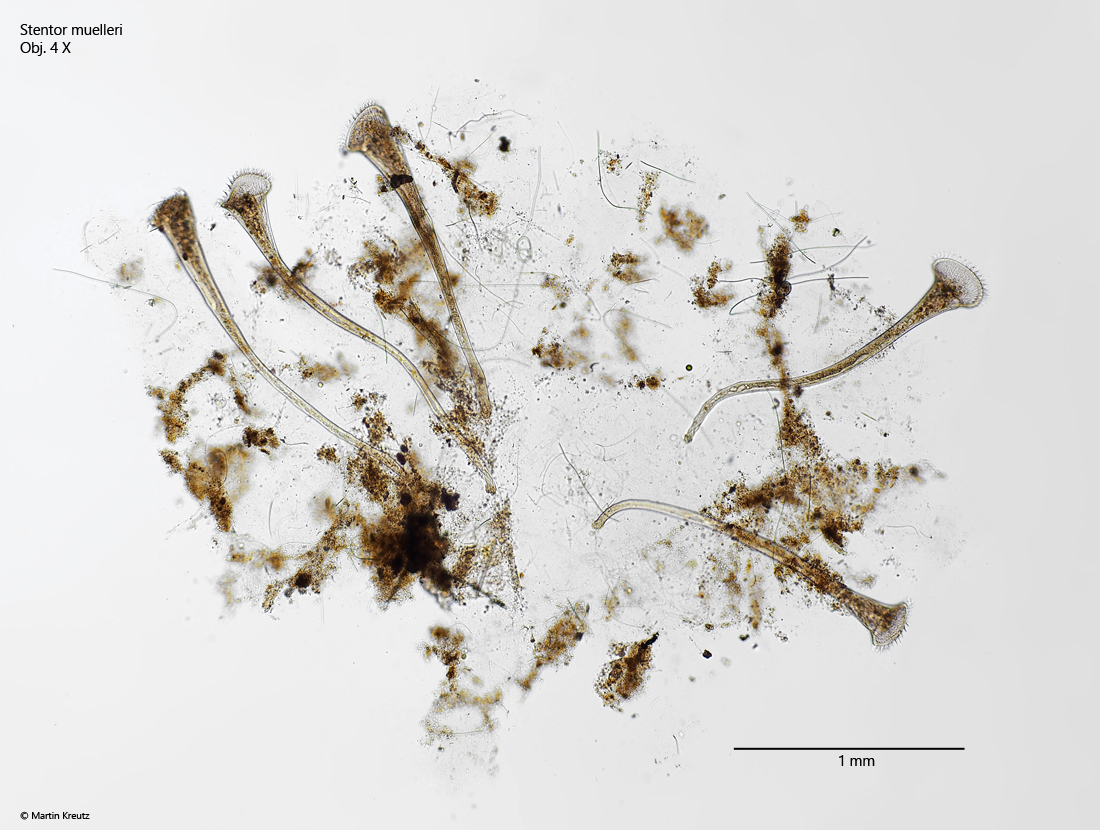
Fig. 1:Stentor muelleri. L = 1100–1350 µm. Some specimen settled in a detritus flake. Obj. 4 X.
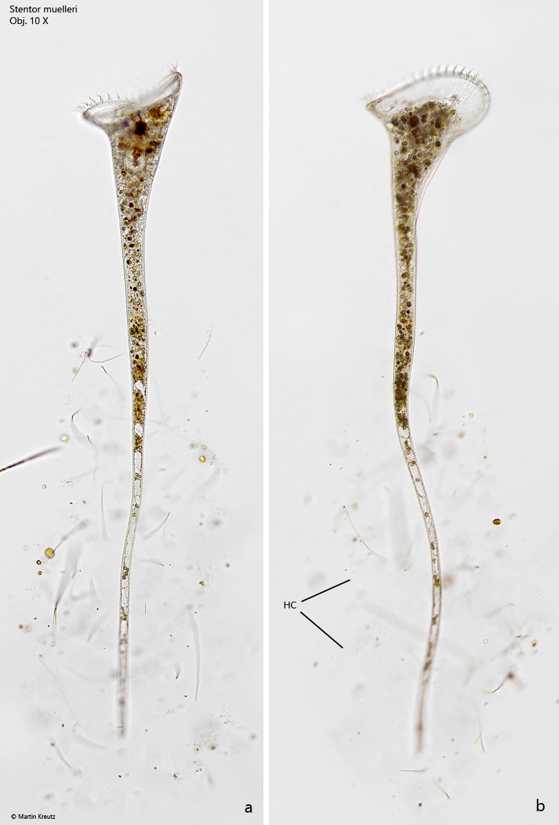
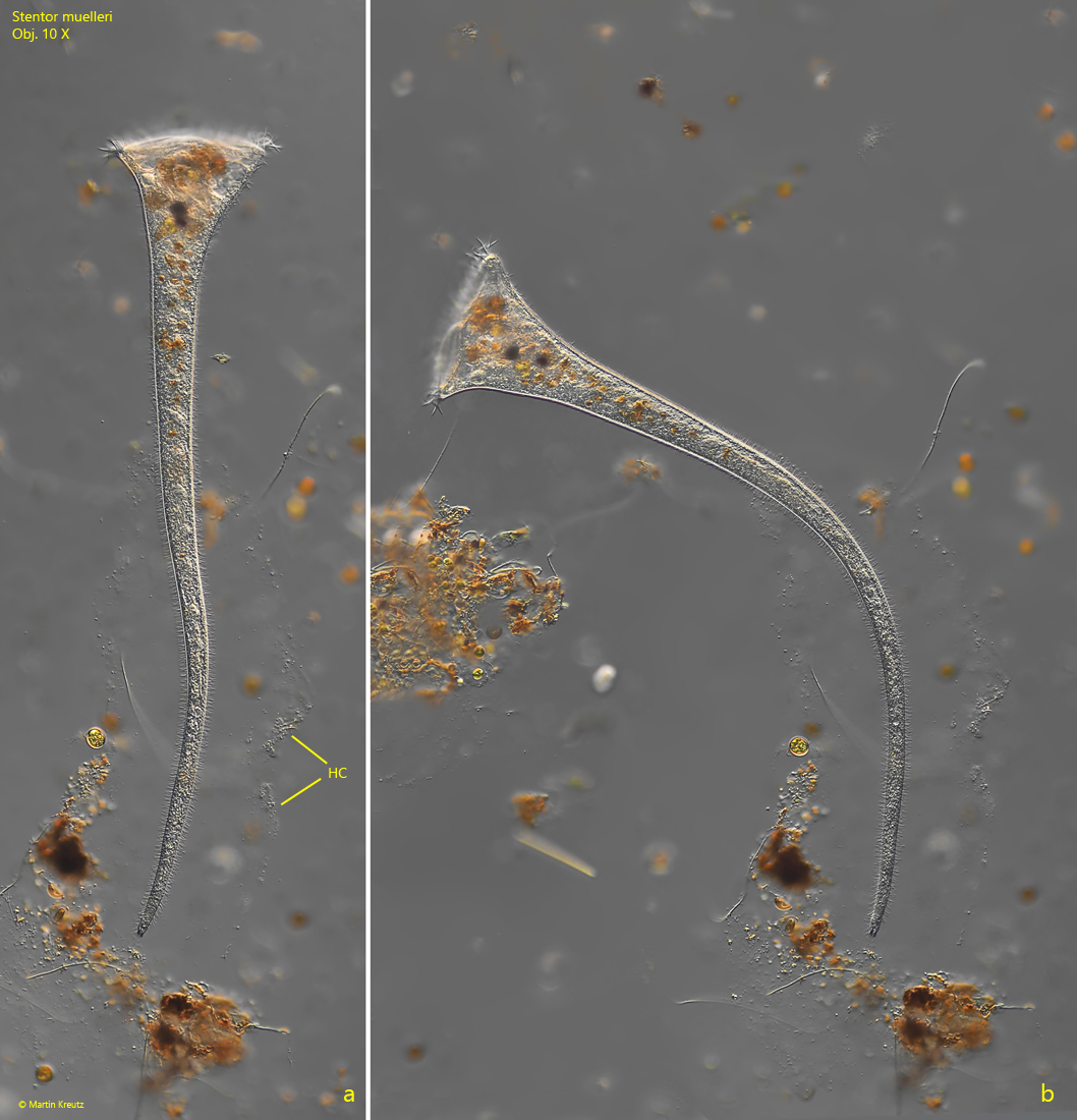
Fig. 2 a-b:Stentor muelleri. L = 1450–1560 µm. Two fully elongated specimens. Note the hyaline case (HC) of the specimens, only visible due to attached particles and algae. Obj. 10 X.
Fig. 3 a-b:Stentor muelleri. L = 1060 µm. A further specimen in a hyaline case (HC) visible due to attached bacteria. Obj. 10 X.
Fig. 4:Stentor muelleri. L = 1060 µm. The anterior half of the specimen as shown in fig. 3 a-b. Note the nodules of the moniliform macronucleus. Obj. 20 X.
Fig. 5:Stentor muelleri. The nodules of the moniliform macronucleus (Ma) with the adjacent micronuclei (Mi) in a strongly squashed specimen. Obj. 100 X.