Stentor roeselii (Ehrenberg, 1835)

Most likely ID: n.a.


Synonym: n.a.


Sampling location: Pond of the convent Hegne, Simmelried


Phylogenetic tree: Stentor roeselii


Diagnosis: 

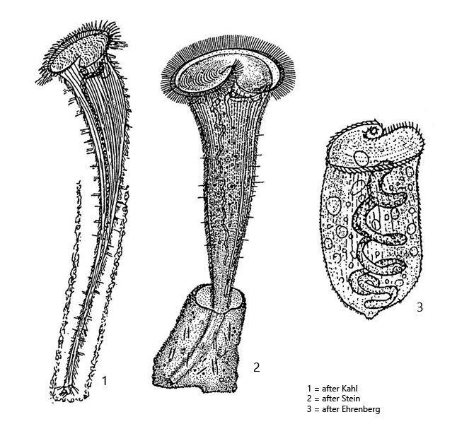
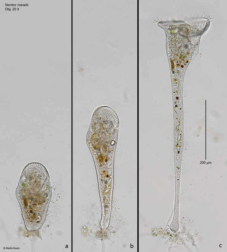
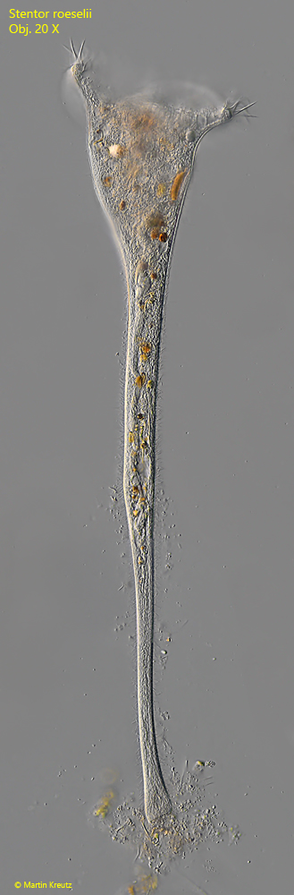
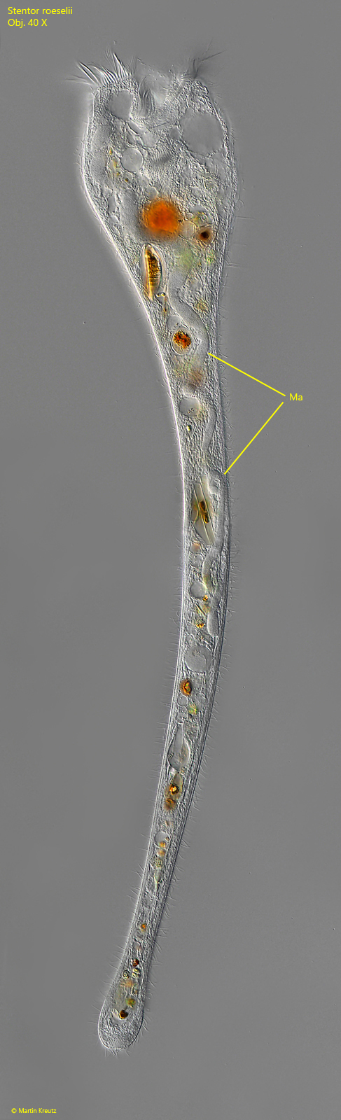
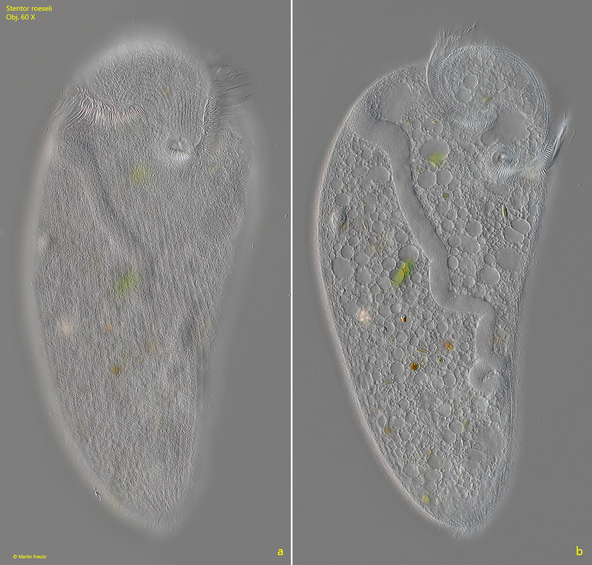
  • body elongated trumpet-shaped, contracted ellipsoid to club-shaped

  • appears yellowish or brownish, no symbiotic algae

  • length 500–1200 µm, sometimes up to 3000 µm (of elongated specimens)

  • adoral membranelle running in clockwise to oral funnel

  • attached with thigmotactic cilia to the substrate

  • sometimes in a hyaline case, 200–300 µm long

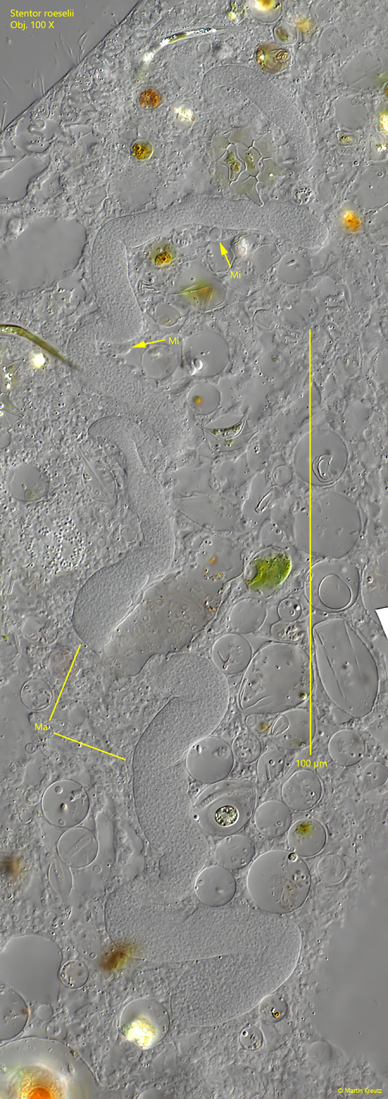
  • macronucleus vermiform, along longitudinal axis of cell

  • 7–20 spherical micronuclei adjacent to the macronucleus

  • contractile vacuole on left wall of oral funnel with one collecting duct reaching posteriorly

Stentor-roeselii
Stentor roeselii

At low magnification, Stentor roeselii resembles Stentor muelleri, which also appears yellowish or brownish. However, Stentor roeselii is about 30 % smaller than Stentor muelleri and appears somewhat plumper when fully elongated (s. fig. 1 c). The decisive distinguishing feature, however, is the shape of the macronucleus. In Stentor roesilii it is vermiform shaped, whereas in Stentor muelleri it is monoliform and resembles a pearl necklace. The shape of the macronucleus can be easily recognized even in slightly squashed specimens (s. figs. 2 and 3 b). In contrast, the small micronuclei, which are attached to the macronucleus, can only be recognized in strongly squashed specimens (s. fig. 5).

Stentor-roeselii

Fig. 1 a-c: Stentor roeselii. L = 710 µm (of elongated specimen). A contracted (a) and fully elongated specimen in brightfield illumination. Obj. 20 X.

Stentor-roeselii

Fig. 2: Stentor roeselii. L = 710 µm. The same specimen as shown in fig. 1 a-c in DIC. Obj. 20 X.

Stentor-roeselii

Fig. 3: Stentor roeselii. L = 730 µm. A slightly squashed specimen. Note the vermiform macronucleus (Ma) arranged longitudinally in the cell. Obj. 40 X.

Stentor-roeseli

Fig. 4 a-b: Stentor roeselii. Focal plane on the pellicle with striation of colorless granules (a) and on the vermiform macronucleus (b, Ma) in a slightly squashed specimen. Obj. 60 X.

Stentor-roeselii

Fig. 5: Stentor roeselii. The vermiform macronucleus (Ma) in a strongly squashed specimen. Two micronuclei (Mi) are in the focal plane. Obj. 100 X.