inhabits a tube-shaped, flexible lorica, about 500 µm long
adoral zone about half of body length
the two apical membranelles of adoral zone are antennae shaped
undulating membranell running parallel to adoral zone
dorsal row of 15 µm long cilia up to cell equator
four frontal cirri
four rows of longitudinal rows of cirri, running spirally in counterclockwise direction
two macronuclei each with an adjacent micronucleus
one contractile vacuole shortly below cytostome on left side
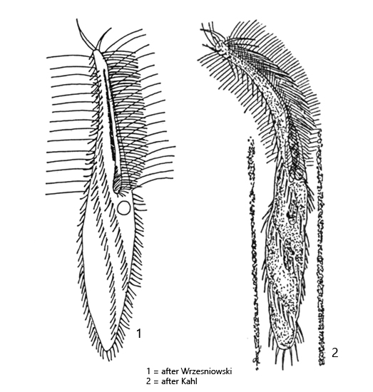
Stichotricha aculeata
I find the hypotrichous ciliate Stichotricha aculeata very frequently in some of my localities. In fresh samples, however, the loricae with the ciliates are difficult to detect as they are often mixed with detritus. It is therefore necessary to leave the samples for 1-2 weeks to allow individuals to colonize the walls of the jar. Stichotricha aculeata also likes to settle on the floating coverslip.
In my sampling sites, Stichotricha aculeata occurs together with the similar species Stichotricha secunda, which, however, has symbiotic algae and can therefore be reliably distinguished from Stichotricha aculeata. Interestingly, Kahl (1932) confused Stichotricha aculeata and Stichotricha secunda, which must have been an absolute exception. As a result, many misidentifications have been made. I base my identification on the descriptions by Foissner et al. (1991).
The characteristics of the individuals of my poulation of Stichotricha aculeata completely agree with the description of Foissner et al. except for the size. The specimens I examined were between 150–240 µm long and sometimes reached twice the length given by Foissner et al. In the literature there are only few data on the length of Stichotricha aculeata. Often the lengths given by earlier authors were adopted. On the Internet, however, you can also find specimens up to 290 µm in length. I therefore consider it likely that the size of my specimens is within the variance of this species.
Fig. 1:Stichotricha aculeata. A colony of about 100 specimens that have settled on a detritus flake. Obj. 10 X.
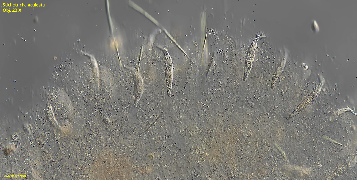
Fig. 2:Stichotricha aculeata. A section of the colony as shown in fig. 1. Obj. 20 X.
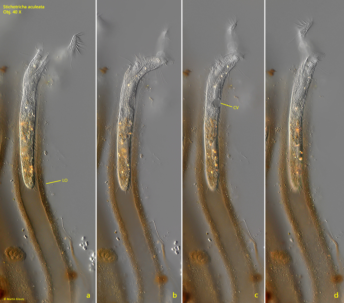
Fig. 3 a-d:Stichotricha aculeata. L = 234 µm. An elongated specimen in the brownish colored lorica (LO). Note the spirally twisted adoral zone. CV = contractile vacuole. Obj. 40 X.
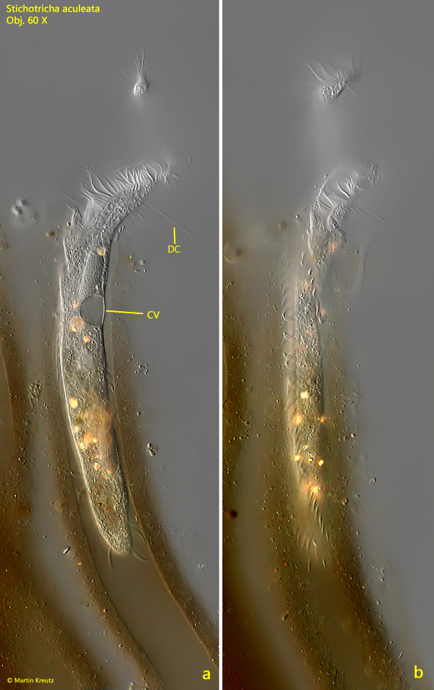
Fig. 4 a-b:Stichotricha aculeata. L = 234 µm. The specimen as shown in fig. 3 a-d in detail. CV = contractile vacuole, DC = dorsal elongated. Obj. 60 X.
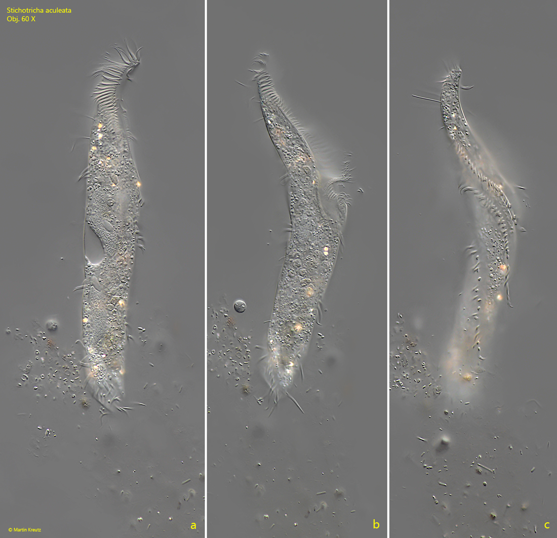
Fig. 5 a-c:Stichotricha aculeata. L = 164 µm. A second specimen in an almost colorless, delicate lorica. Obj. 60 X.
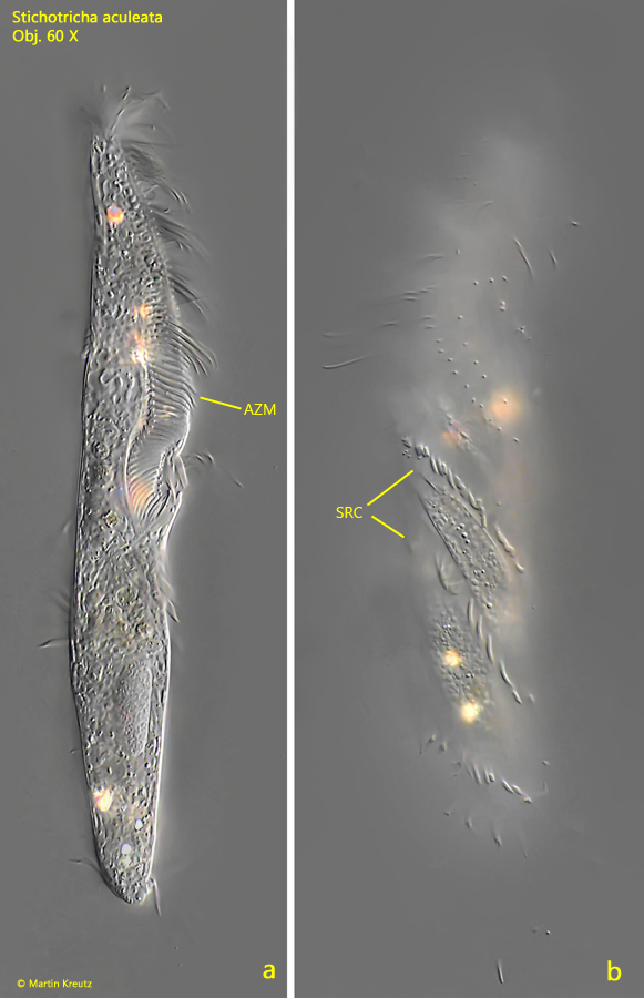
Fig. 6 a-b:Stichotricha aculeata. L = 172 µm. Two focal planes of a freely swimming specimen. AZM = adoral zone of membranelles, SRC = spirally rows of cirri. Obj. 60 X.
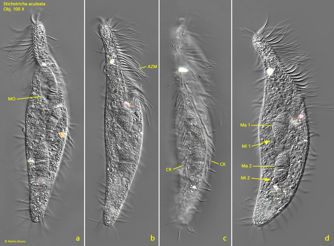
Fig. 7 a-d:Stichotricha aculeata. L = 165 µm. Different focal planes of a slightly squashed specimen. AZM = adoral zone of membranelles; CR = spirally rows of cirri; Ma 1, Ma 2 = macronuclei; Mi 1, Mi 2 = micronuclei. Obj. 100 X.
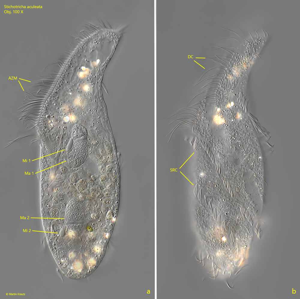
Fig. 8 a-b:Stichotricha aculeata. L = 156 µm. Two focal planes of a second, squashed specimen. DC = dorsal row of elongated cilia; Ma 1, Ma 2 = macronuclei; Mi 1, Mi 2 = micronuclei; SRC = spirally rows of cilia. Obj. 100 X.