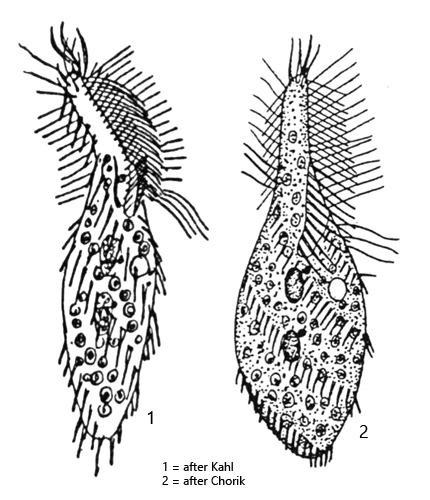
the two apical membranelles of adoral zone are antennae shaped
undulating membranell running parallel to adoral zone
dorsal row of elongated cilia in the area of adoral zone
spirally rows of cirri in counterclockwise direction
two macronuclei each with an adjacent micronucleus
one contractile vacuole shortly below cytostome on left side
Stichotricha secunda
The hypotrichous ciliate Stichotricha secunda was very rare in the Simmelried until around the year 2010, but then became increasingly common. Especially in the last 5 years it became more common than the similar species Stichotricha aculeata.
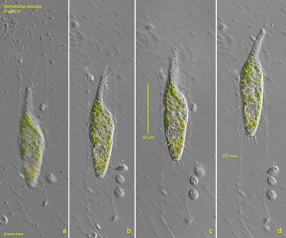
Stichotricha secunda builds and lives in a tubular lorica. According to my observations, however, it is at most 2-3 times as long as the ciliate, i.e. 200-300 µm. The loricae are almost always solitary. Larger groups of more than 3 individuals are rarely found. However, Stichotricha secunda is usually found swimming freely, as the lorica is abandoned at the slightest disturbance. In addition, the loricae are hardly recognizable in fresh samples as they are very delicate, colourless and transparent. The floating coverslip has proven to be the most suitable method for observing Stichotricha secunda in the lorica. It is colonized by Stichotricha secunda after just a few days.
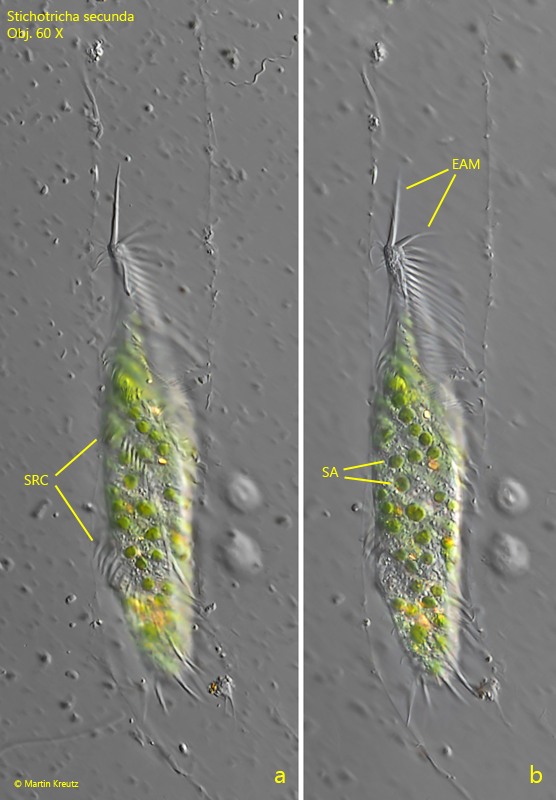
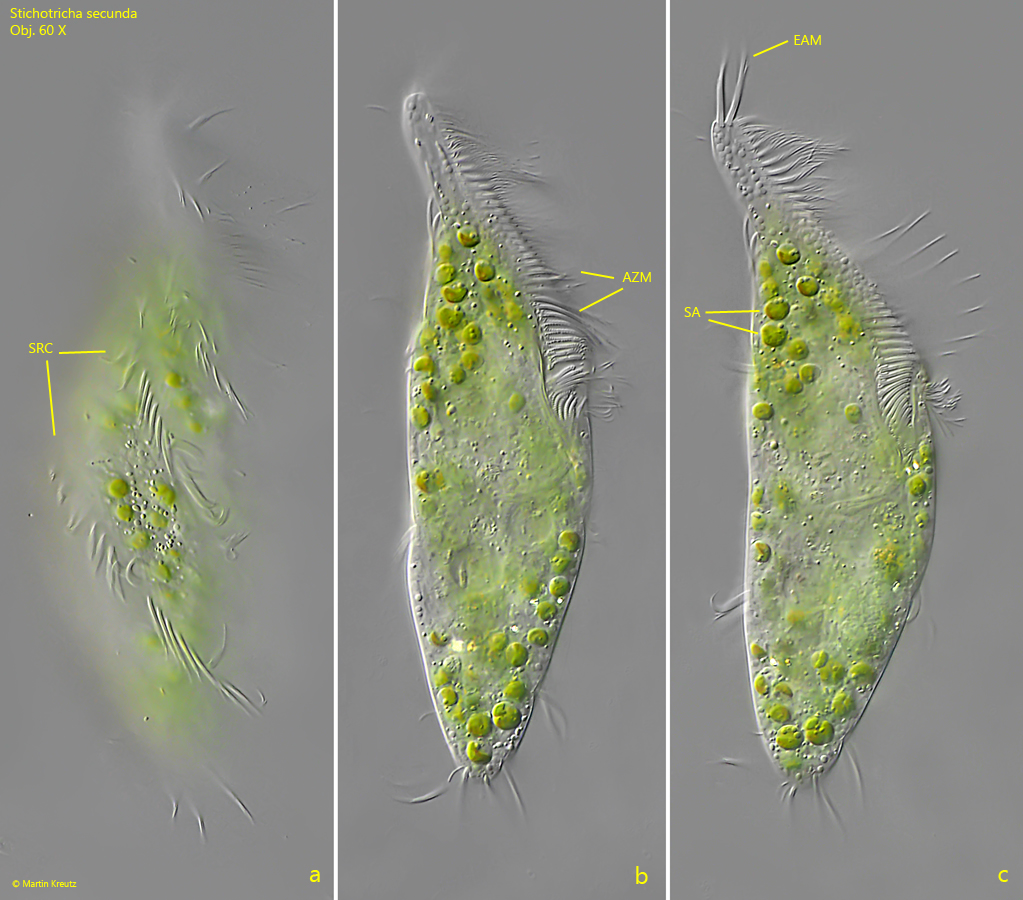
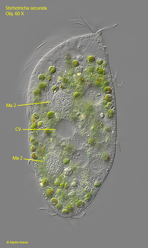
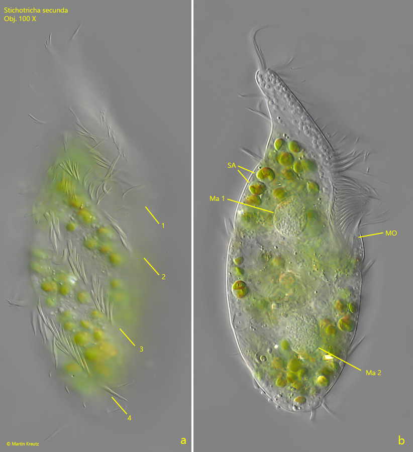
The descriptions of Stichotricha secunda by earlier authors are very brief and incomplete. Foissner et al. mention that the number of spiral rows of cirri running longitudinally across the body is not exactly known. According to my observations there are 4 of these spiral rows (s. fig. 5 a), the same number as in the similar species Stichotricha aculeata. In contrast to Stichotricha aculeata, the adoral zone of Stichotricha secunda is not spirally twisted in elongated specimens (s. figs. 1 c and 1 d). The two apical membranelles of the adoral zone are transformed into two antenna-shaped cirrus with a length of about 15-18 µm (s. figs. 2 b and 3 c). The cytoplasm is filled with about 100 symbiotic algae, which have a diameter of 3–5 µm and a cup-shaped chloroplast. A pyrenoid is not present. They are not of the Chlorella type. Crushed specimens burst very quickly and denature strongly. Therefore, I have only been able to identify the two macronuclei (s. figs. 4 and 5 b), but not the two attached micronuclei. The specimens in my population were 100–140 µm long. The adoral zone reached almost exactly 50 % of the body length in elongated specimens (s. fig. 1 c).
Fig. 1 a-d:Stichotricha secunda. L = 110 µm. A specimen moving in the tube-shaped lorica (LO). Obj. 40 X.
Fig. 2 a-b:Stichotricha secunda. L = 110 µm. The same specimen as shown in fig. 1 a-d at higher magnification. Note the two elongated, antennae-shaped adorale membranelles (EAM). SA = symbiotic algae, SRC = spirally rows of cirri. Obj. 60 X.
Fig. 3 a-c:Stichotricha secunda. L = 128 µm. Different focal planes of a slightly squashed specimen. AZM = adoral zone of membranlles, EAM = elongated adoral membranelles, SA = symbiotic algae, SRC = spirally rows of cirri. Obj. 60 X.
Fig. 4:Stichotricha secunda. The squashed specimen as shown in fig. 3 a-c. The two macronuclei are visible (Ma 1, Ma 2) but not the adjacent micronuclei. Obj. 60 X
Fig. 5 a-b:Stichotricha secunda. L = 105 µm. A second, slightly squashed specimen. Note the 4 spirally rows of cirri (1–4). Ma 1, Ma 2 = macronuclei; MO = mouth opening; SA = symbiotic algae. Obj. 100 X
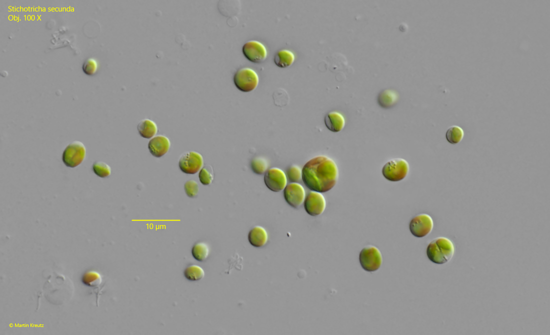
Fig. 6:Stichotricha secunda. The symbiotic algae released from a strongly squashed specimen. The algae cells have a diameter of 3–5 µm (dividing cells excluded) and a cup-shaped chloroplast. A pyrenoid is absent and they are not from the Chlorella type. Obj. 100 X