adoral zone arises dorsally, runs almost equatorially to ventral side where it bends posteriorly
posterior end narrowly rounded or pointed, not tail-like
macronucleus large, globular with adjacent spherical micronucleus
prominent fringe of extrusomes (about 3 µm long) beneath pellicle
contractile vacuole subterminal
tuft of elongated caudal cilia at posterior end
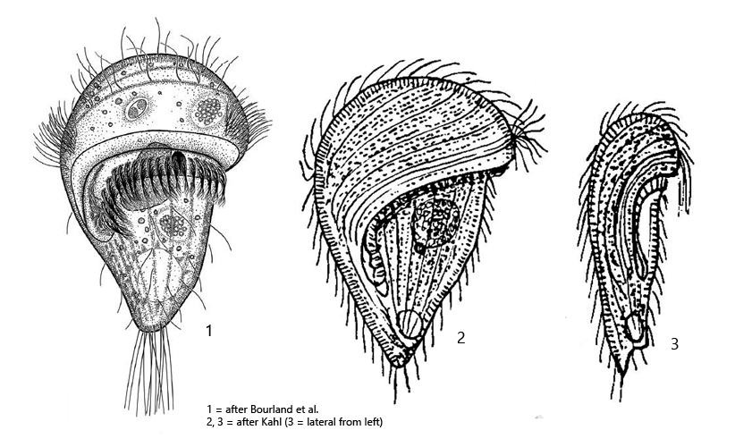
Urostomides bacillatus
Urostomides bacillatus was first described in 1894 by Levander as Metopus bacillatus. In 2017 the species was redescribed by Bourland et al. who transferred the species to the genus Urostomides.
Urostomides bacillatus is very similar to Urostomides striatus. However, Urostomides striatusis smaller (< 100 µm), has a distinctly tail-like extended posterior end and is also much more dorso-ventrally flattened.
I find Urostomides bacillatus regularly and frequently in the mud zone. The species looks somewhat mushroom shaped when free swimming. Close examination is complicated by coverglass sensitivity. However, I was able to determine that a few specimens squeeze easily under the coverslip, while most specimens denature before. The reasons for this different behavior is not known to me.
Kahl mentions that the species is very variable. I can confirm this by the variability of the posterior half of the body. Some specimens were shaped like an ice cream cone, while in other specimens the posterior end appeared distinctly tapered, but this also depended on the degree of filling of the contractile vacuole. This makes it difficult to distinguish Urostomides bacillatus from Urostomides striatus.
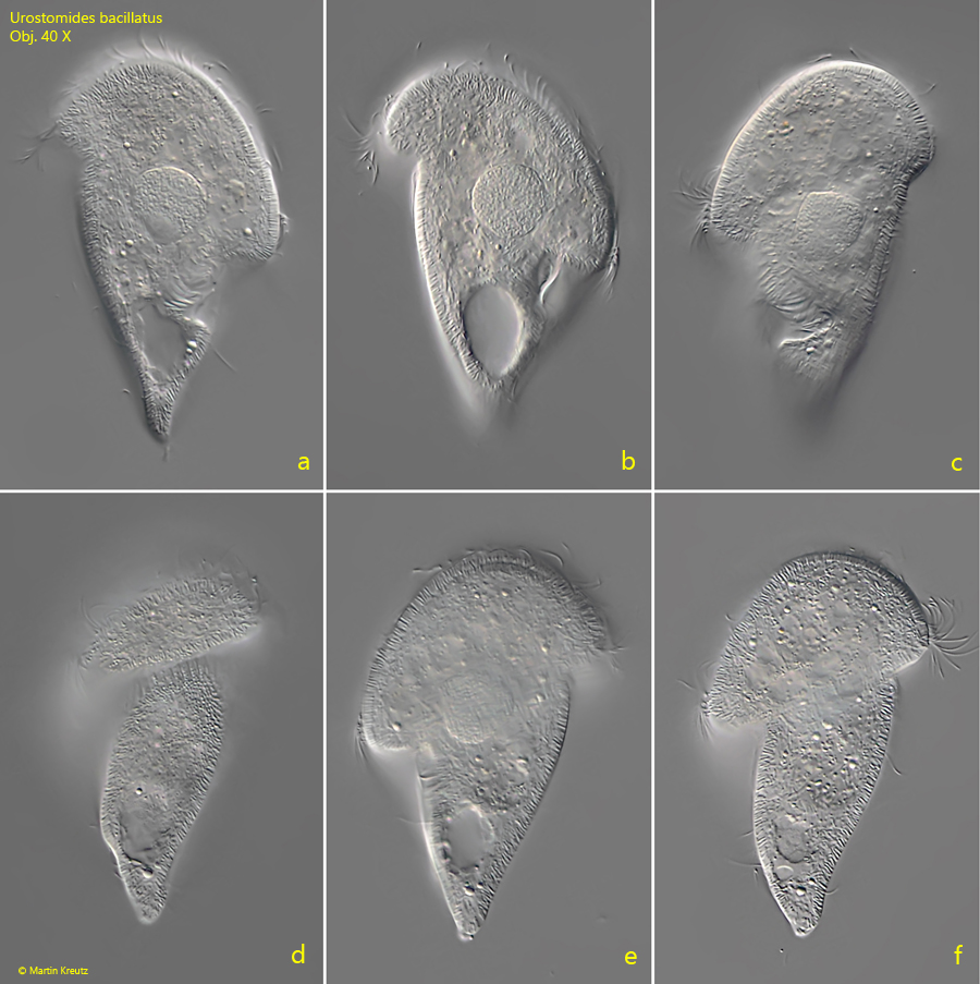
Fig. 1 a-f:Urostomides bacillatus. L = 100 µm. A freely swimming specimen from dorsal (a, b), ventral (c) and from right (d, e, f). Obj. 40 X.
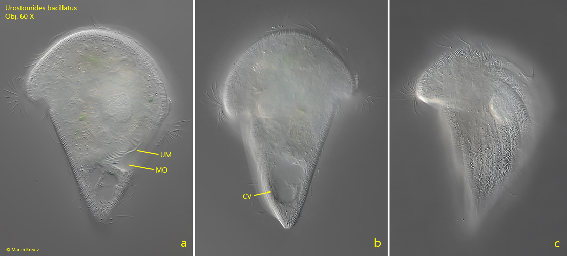
Fig. 2 a-c:Urostomides bacillatus. L = 114 µm. A second freely swimming specimen from dorsal (a, c) and from left (b). CV = contractile vacuole, MO = mouth opening, UM = undulating membrane. Obj. 60 X.
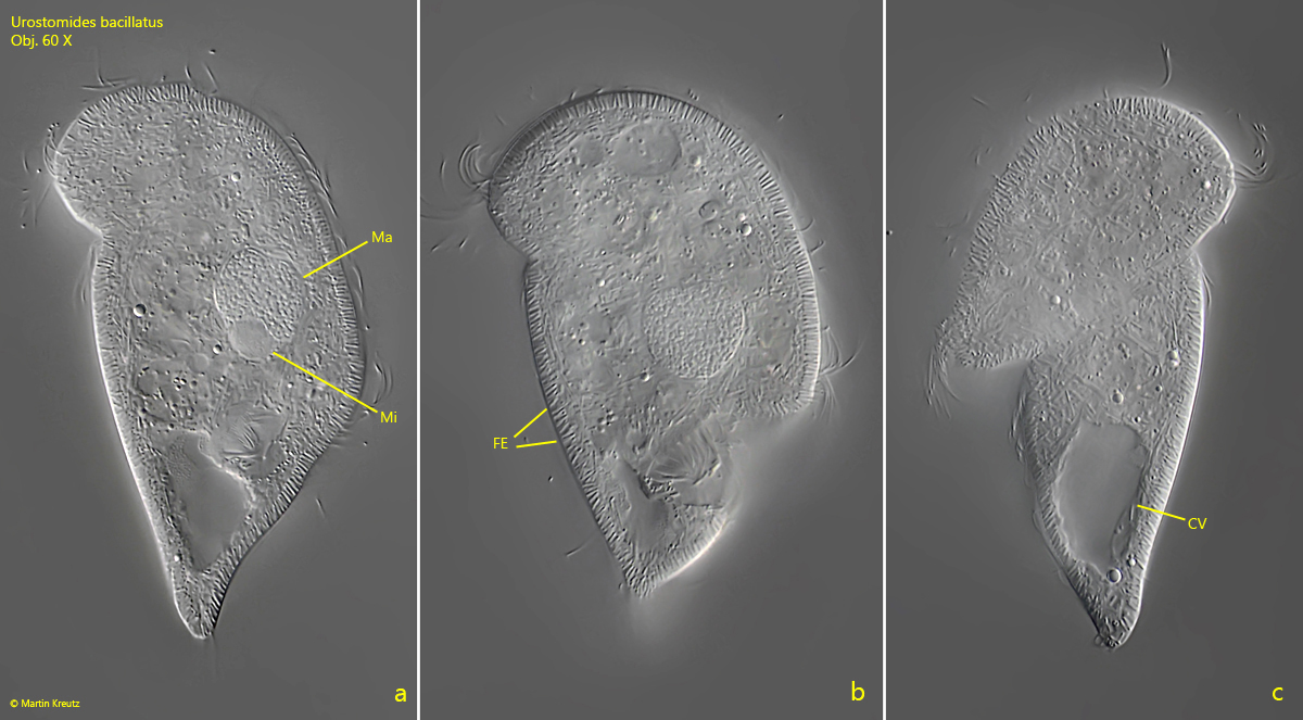
Fig. 3 a-c:Urostomides bacillatus. L = 100 µm. A third freely swimming specimen from dorsal (a, c) and from right (c). CV = contractile vacuole, FE = fringe of extrusomes, Ma = macronucleus, Mi = micronucleus. Obj. 60 X.
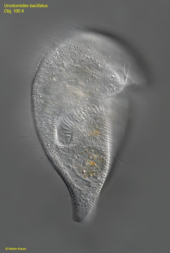
Fig. 4:Urostomides bacillatus. L = 124 µm. A freely swimming specimen from ventral. Obj. 100 X.
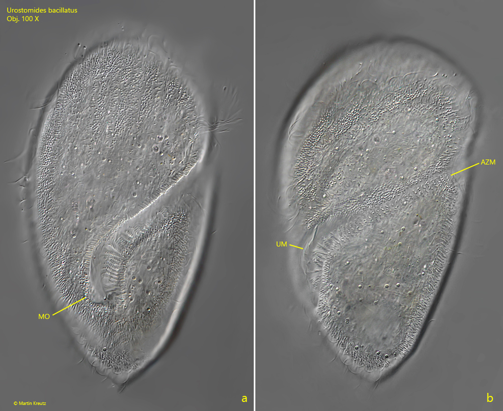
Fig. 5 a-b:Urostomides bacillatus. L = 128 µm. A slightly squashed specimen from ventral. AZM = adoral zone of membranelles, MO = mouth opening, UM = undulating membrane. Obj. 100 X.
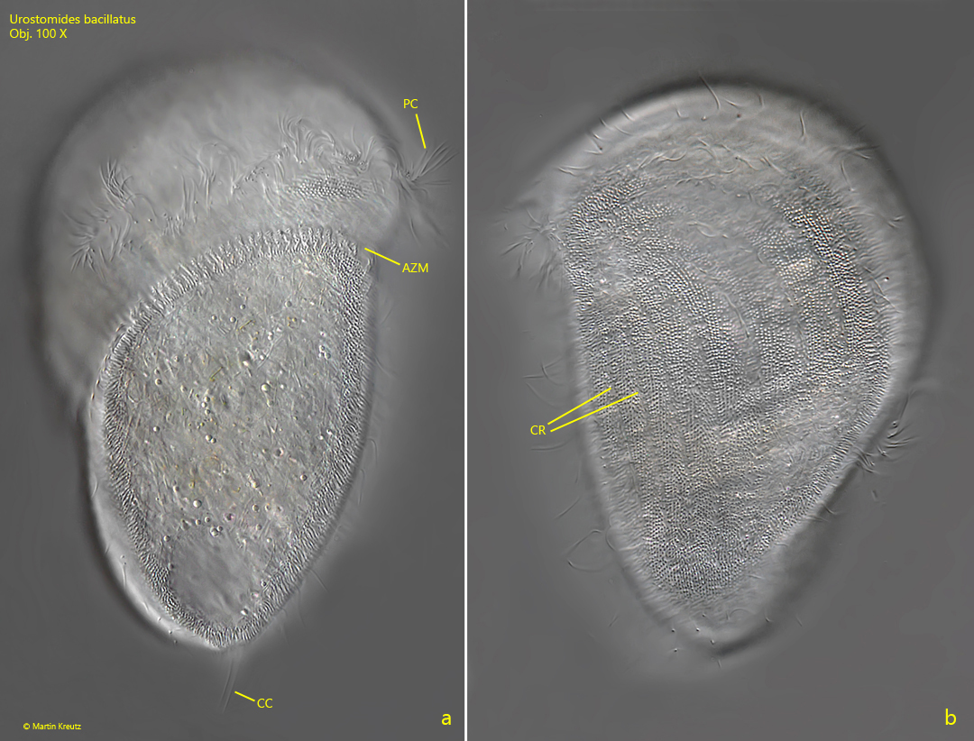
Fig. 6 a-b:Urostomides bacillatus. L = 128 µm. The same specimen shown in fig. 5 a-b from right (a) and from dorsal (b). Note the distinct striation of the pellicle by the ciliary rows (CR). In the space between the ciliary rows the exutrusomes are visible as bright dots. AZM = adoral zone of membranelles, CC = caudal cilia, PC = perizonal cilia. Obj. 100 X.