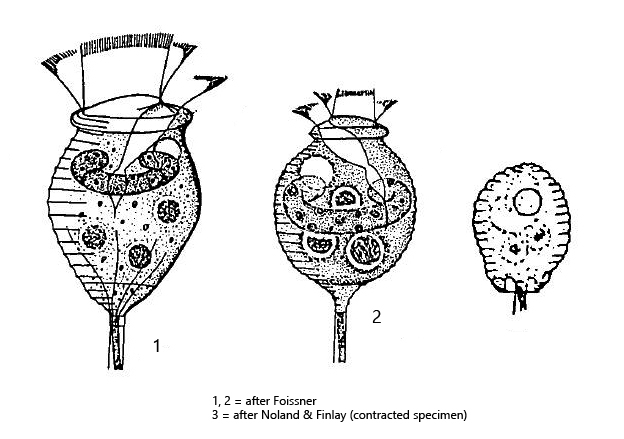
macronucleus horseshoe-shaped, located in anterior half
pellicle is widely striped, 13–30 horizontal stripes
stalk contracts in spirals
solitary
Vorticella aquadulcis
I found Vorticella aquadulcis in 1998 in Hagstaffel pond attached to colonies of the planktonic cyanobacterium Anabaena flos-aquae and 2023 on coverslips floating on samples from Simmelried . Vorticella aquadulcis can be easily recognized by the widely spaced striation of the pellicle and the horseshoe-shaped macronucleus, which is located in the anterior half of the cell. The species occurs exclusively solitary and does not form pseudocolonies as do many other species of the genus Vorticella. Many synonymous species are grouped under Vorticella aquadulcis, which is why it is also called the “Vorticella aquadulcis complex” (s. Synonyms above).
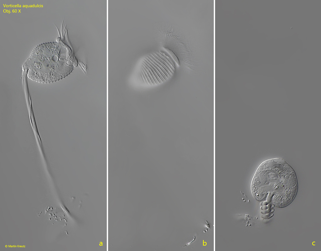
Fig. 1 a-c: Vorticella aquadulcis. L = 32 µm. Two focal planes of a fully extended specimen (a, b) and a fully contracted specimen (c). Note the widely spaced striation of the pellicle (b). Obj. 60 X.
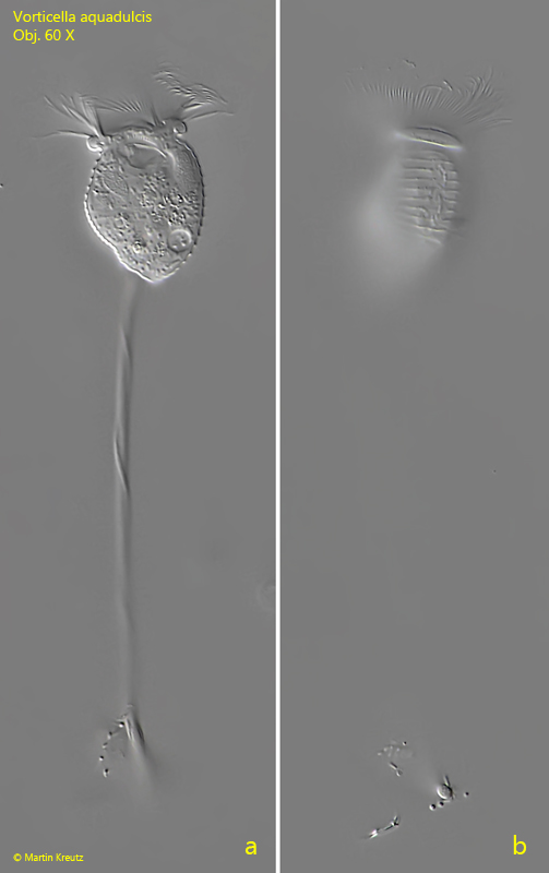
Fig. 2 a-b: Vorticella aquadulcis. L = 31 µm. Two focal planes of a seceond, fully extended specimen. Obj. 60 X.
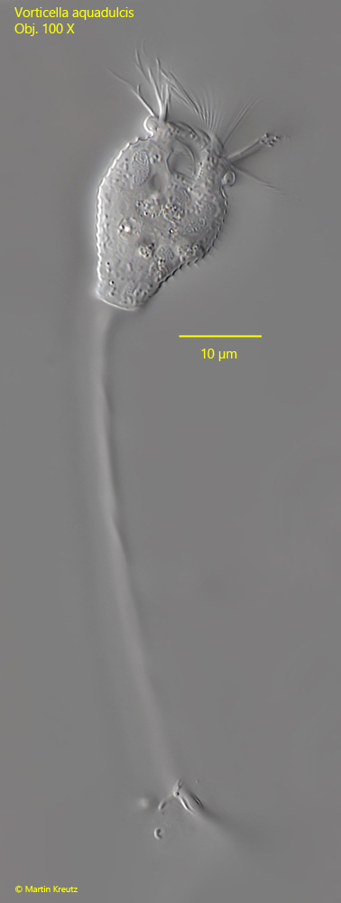
Fig. 3: Vorticella aquadulcis. L = 33 µm. A fully extended specimen in detail. Obj. 100 X.
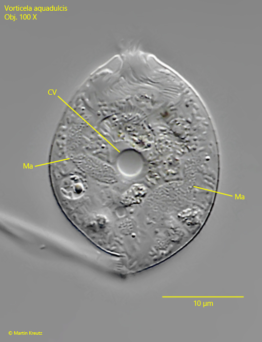
Fig. 4: Vorticella aquadulcis. A strongly squashed specimen to visualize the single contractile vacuole (CV) and the horseshoe-shaped macronucleus (Ma). Obj. 100 X.