body lanceolate, left side flat, right side convex
length 30–60 µm, width 10–15 µm
mouth provided with extrusomes
mouth curved to the left side at the apical end
extrusomes straight, 2–3 µm long
two ellipsoidal macronuclei, one spherical micronucleus between them
right side with 2 rows of cilia (without perioral row of cilia)
left side with 2 rows of short, bristle-like cilia
dorsal brush a short row of paired bristles
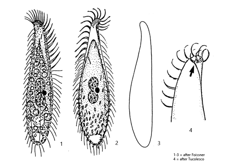
Acinera uncinata
I usually find Acinera uncinata on the floating coverslip. The specimens are difficult to find in fresh samples because they usually adhere to detritus flakes.
Acinera uncinata resembles Litonotus lamella at first sight. However, Litonotus lamella is larger, has curved extrusomes which are also present on the posterior end and the ciliated right side has 4–7 rows of cilia. In Acinera uncinata, only 3 rows of cilia are recognizable on the right side, one of which is the perioral row, which can be recognized by the fact that it consists of more densely arranged cilia (s. fig. 1 c). The extrusomes of Acinera uncinata are straight rods of 2–3 µm in length and are located only at the mouth opening. Another important feature is the apical end, which is folded over to the left (unciliated) side (s. drawing 4 , above). This is only clearly visible in dorsal or ventral view (s. fig. 1 d). On the floating coverslip, the specimens are almost exclusively with the (ciliated) right side facing the objective, on which they slide along the coverslip. Therefore it is difficult to examine the left side, which has only 2 rows of short bristles.
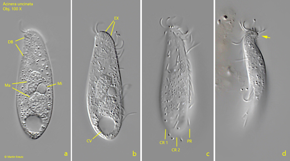
Fig. 1 a-d:Acinera uncinata. L = 31 µm. A freely swimming specimen from right (a-c) and from ventral (d). Note the apical end curved to the left side (arrow). The ciliation of the right side is consisting of 2 rows of cilia (CR 1, CR 2) and a perioral row of cilia (PR). The cilia of the perioral row are closer together. CV = contractile vacuole, DB = dorsal brush, EX = extrusomes, Ma = macronuclei, Mi = micronucleus. Obj. 100 X.
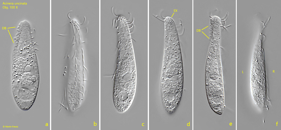
Fig. 2 a-f:Acinera uncinata. L = 36 µm. A second freely swimming specimen from right (a-d) and from dorsal (e, f). DB = dorsal brush, EX = extrusomes, L = left side, R = right side. Obj. 100 X.