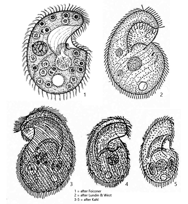
macronucleus globular to slighty ellipsoid, nucleolus reticulate
one micronucleus adjacent to macronucleus, about 3 x 1.5 µm
mouth opening with a right and left field of polykineties
extrusomes inconspicuous, 0.8–1 µm long
ciliary rows consiting of paired cilia
contractile vacuole almost terminal
no caudal cilia
Bresslaua vorax
I found Bresslaua vorax in moss samples that came mainly from trees and walls. If the moos is poured over with rainwater in Petri dishes, large populations of Bresslaua vorax occurred in some of the samples. Bresslaua vorax has so far been found in mosses, hay infusions and soil samples. Limnetic records has not yet been confirmed.
Bresslaua vorax is easily confused with Colpoda cucullus in terms of size and shape. However, the anterior half of Colpoda cucullus is much more broadly rounded, the body is kidney-shaped and the mouth opening is smaller. In addition, Colpoda cucullus is exclusively feeding on bacteria, whereas Bresslaua vorax is a predator of other ciliates (s. fig. 6). In addition, the extrusomes of Colpoda cucullus are larger and comma-shaped.
In my populations, the specimens of Bresslaua vorax were 80 – 95 µm long. I have not found any larger specimens. However, with good nutrition giant forms up to 250 µm were described by Kahl (1931). The specimens in my population mainly preyed mainly Colpoda steinii. Some specimens were completely opaque and deformed due to the high number of ingested ciliates. I found transparent specimens of Bresslaua vorax only in old samples, with a low concentration of prey ciliates.
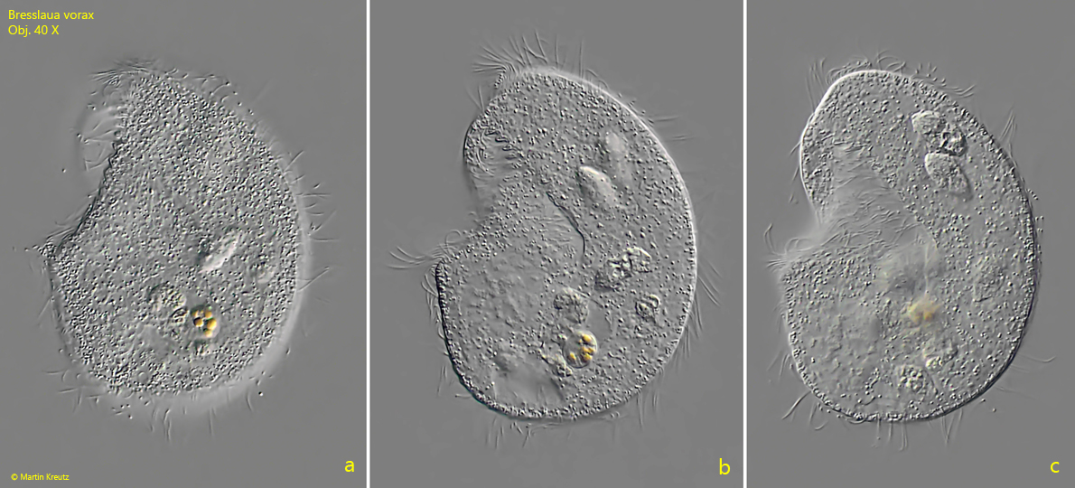
Fig. 1 a-c:Bresslaua vorax. L = 89 µm. A freely swimming specimen from left. Obj. 40 X.
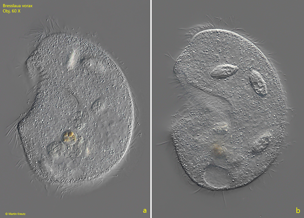
Fig. 2 a-b:Bresslaua vorax. L = 89 µm. The same specimen as shown in fig. 1 a-c at higher magnification. Obj. 60 X.
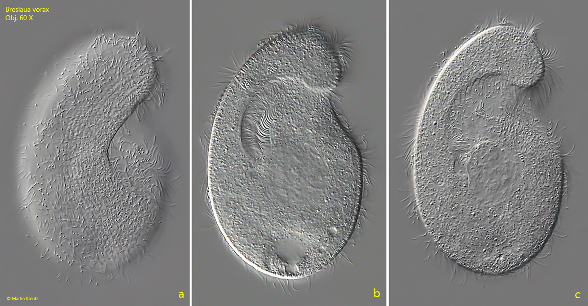
Fig. 3 a-c:Bresslaua vorax. L = 94 µm. A freely swimming specimen from right. Obj. 60 X.
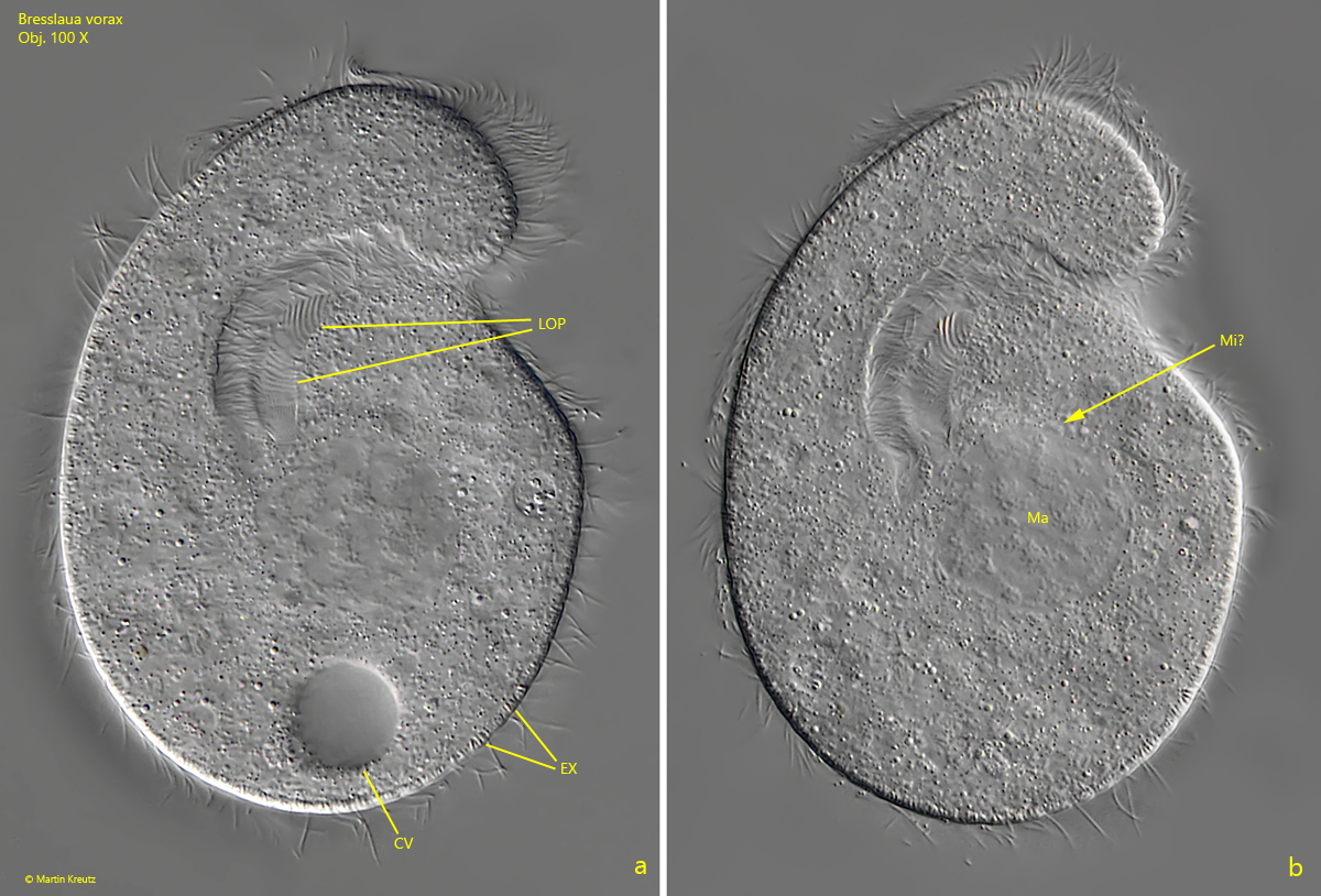
Fig. 4 a-b:Bresslaua vorax. L = 94 µm. Two focal planes of the slighty squashed specimen as shown in fig. 3 a-c. Note the curved oral polykinetid of the left side (LOP). CV = contractile vacuole, EX = extrusomes, Ma = macronucleus with reticulate nucleolus, Mi? = probably the micronucleus. Obj. 100 X
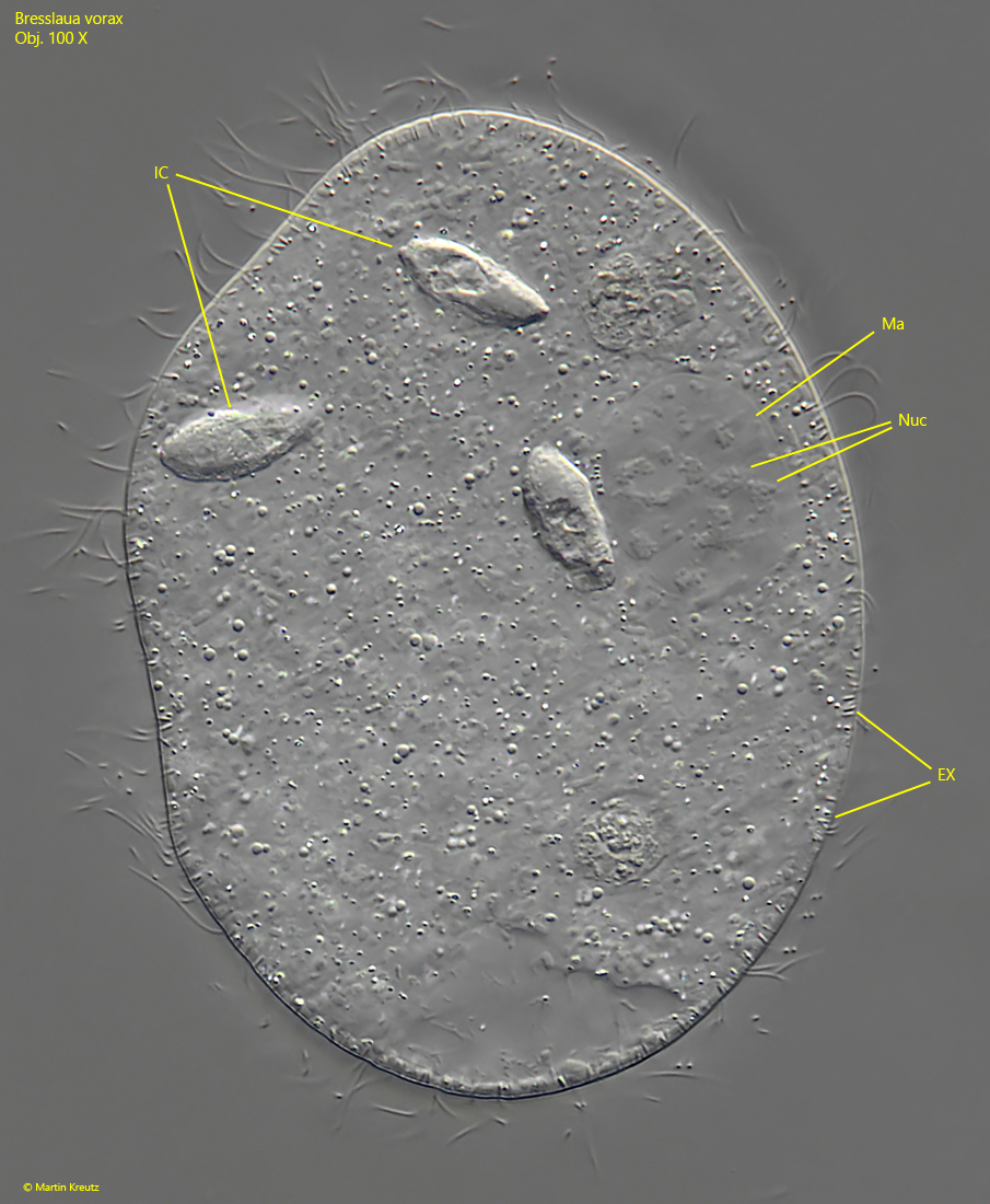
Fig. 5:Bresslaua vorax. A strongly squashed specimen. Note the ingested ciliates (IC). EX = extrusomes, Ma = macronucleus with reticulate nucleolus, Nuc = nucleolus. Obj. 100 X.
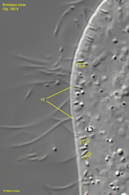
Fig. 6:Bresslaua vorax. The extrusomes (EX) with a length of 1–1.2 µm in detail. Obj. 100 X.
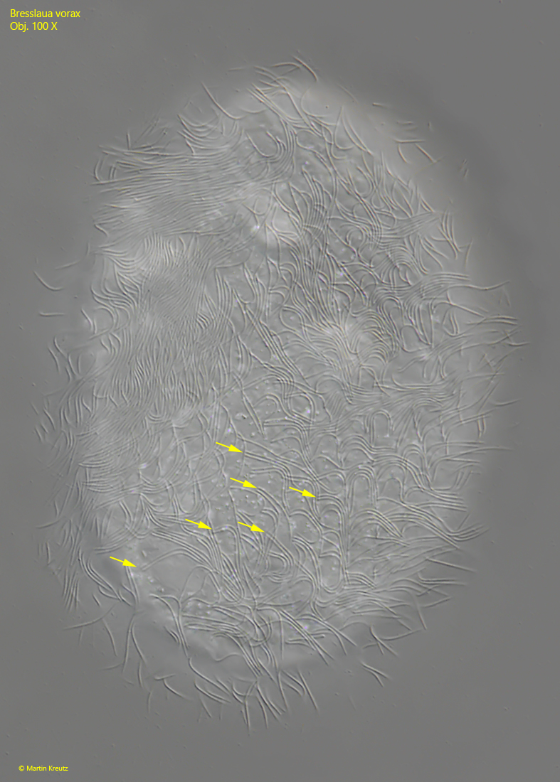
Fig. 7:Bresslaua vorax. Focal plane on the ciliation of a strongly squashed specimen. Note the paired cilia (arrows) of the somatic ciliation. Obj. 100 X.