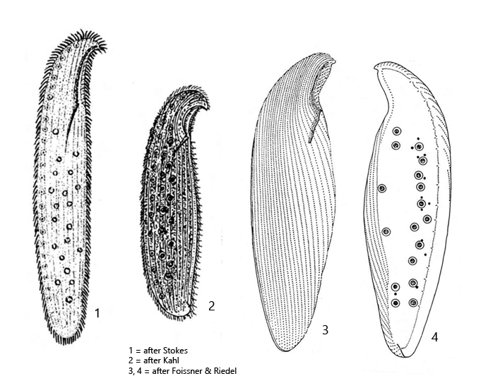
body slender to broadly ellipsoid, anterior end beak-shaped, laterally strongly flattened
length 300–800 µm, width 80–150 µm
3–31 macronuclei (mostly 17) distributed in the cytoplasm in two indistinct rows
2–32 micronuclei (mostly 12), some distributed in the cytoplasm, some adjacent to the macronuclei
no contractile vacuole
a row of about 10–24 Müller vesicles along the dorsal side
cytoplasm mostly strongly vacuolated
pellicle with parallel rows of brownish granules (sometimes colorless)
right side with 25–32 longitudinal rows of paired cilia
left side with only two marginal rows of cilia
oral apparatus immediately behind the beak-shaped anterior end
Loxodes magnus
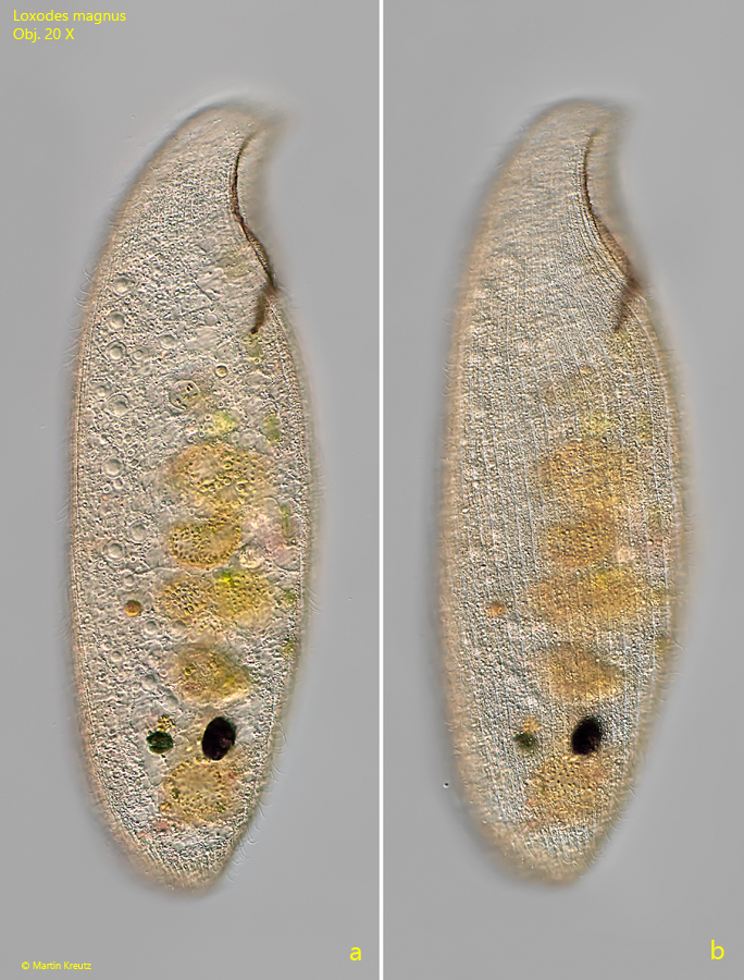
Loxodes magnus is one of the largest and most conspicuous ciliates in the sapropel. I find it in all my sampling sites with a layer of decaying mud. The specimens are mostly brownish or yellow-brown colored due to pigment granules in the pellicle. But I have also found almost colorless specimens (s. fig. 3). Often Loxodes magnus is found together in the samples with the other two species Loxodes rostrum and Loxodes striatus. From the body shape the Loxodes species look similar, by the curved, beak-shaped anterior end. However, Loxodes magnus differs from these two species by its size (mostly > 400 µm) and by a large number of macronuclei (mostly 17) distributed in the cytoplasm (s. figs. 2 and 3). The micronuclei, which are also present in large numbers, are partially adjacent to the macronuclei or are also distributed in the cytoplasm (s. fig. 3). The other two species Loxodes striatus and Loxodes rostrum possess only two macronuclei, which are either widely separated (Loxodes striatus) or close together (Loxodes rostrum). In addition, Loxodes rostrum possesses characteristic symbiotic algae, which are absent in Loxodes magnus.
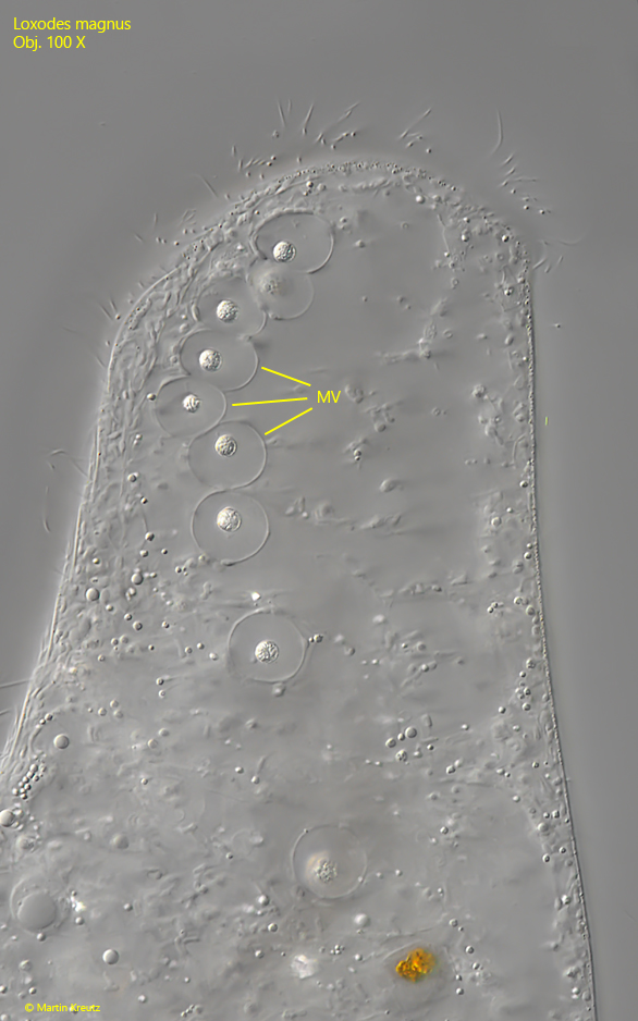
The genus Loxodes has specially constructed Müller vesicles. These are constant vacuoles in which a druse of barium sulfate crystals sits on a plasma stalk (s. also Loxodes striatus). It is assumed that this organelles serves for orientation in the water body, similar to an organ of balance. Loxodes magnus has 10–24 of these organelles, which are arranged in a row on the dorsal side. They can be recognized quite easily by the highly refractive barium sulfate crystals that shine brightly in DIC (s. fig. 4).
Fig. 1 a-b:Loxodes magnus. L = 812 µm. Two focal planes of a freely swimming specimen. Obj. 20 X.
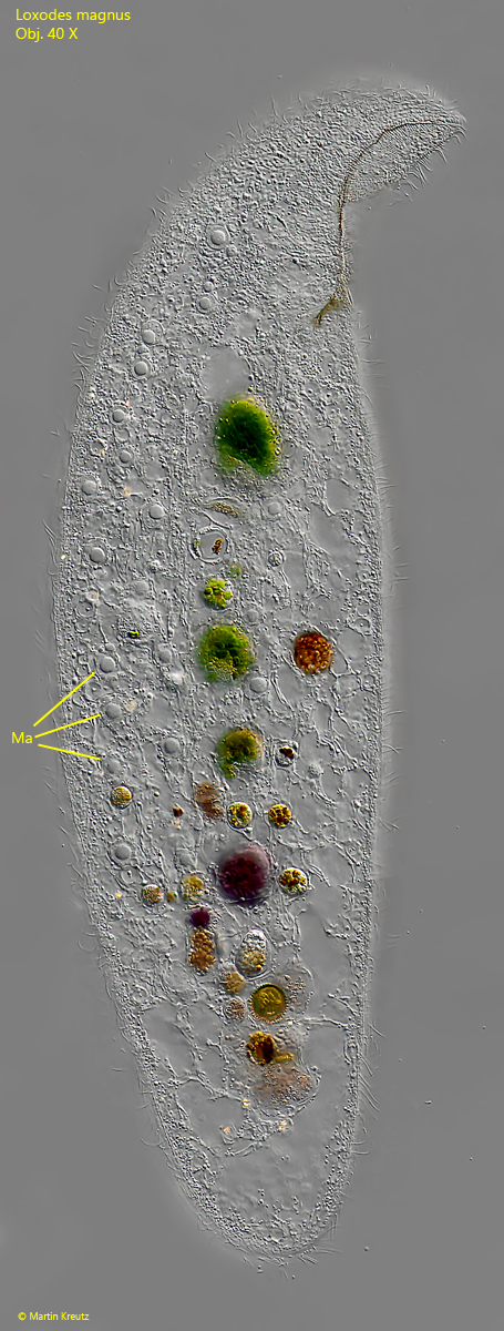
Fig. 2:Loxodes magnus. L = 504 µm. A slightly squashed, transparent specimen. Note the macronuclei (Ma) distributed in the cytoplasm. Obj. 40 X.
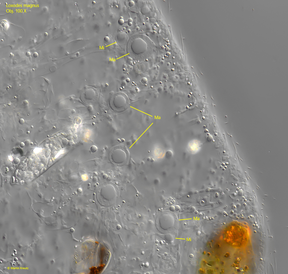
Fig. 3:Loxodes magnus. The macronuclei (Ma) in a strongly squashed specimen. Some of them have micronuclei (Mi) attached. Obj. 100 X.
Fig. 4:Loxodes magnus. The anterior end of a specimen from dorsal. The Müller vesicles (MV) are arranged in a row. Obj. 100 X.
Fig. 5:Loxodes magnus. Two focal planes of the anterior end from right where the mouth opening (MO) is located. The tube covered with brown granules is the pharyngeal basket (PB). Obj. 100 X.
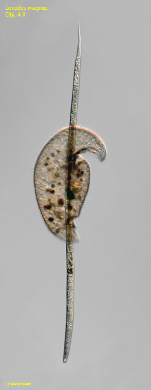
Fig. 6:Loxodes magnus. I found this curious scene in August 1999. A specimen has phagocytized a nematode with a length of 1400 µm. Half of it was already excreted from the cytopyge. The whole prey was carried along during swimming. Obj. 4 X.