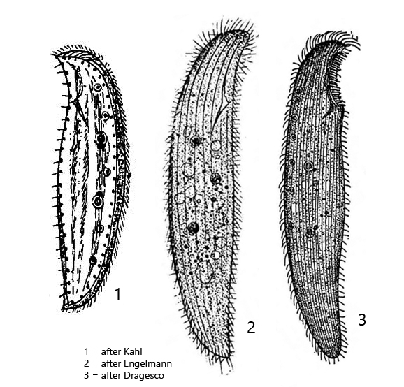
body slender to broadly ellipsoid, anterior end beak-shaped, laterally strongly flattened
length 100–300 µm, width 25–60 µm
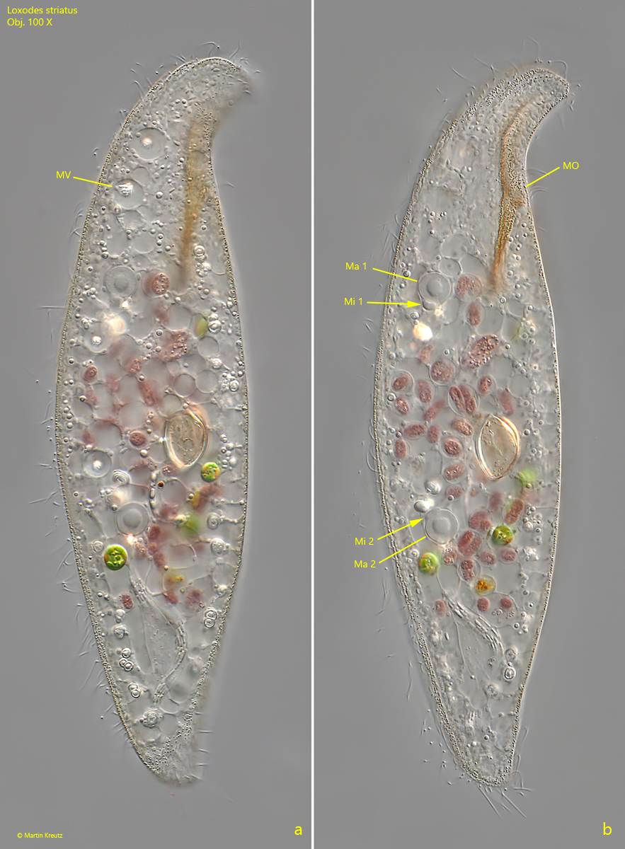
two widely separated macronuclei with one micronucleus each
no contractile vacuole
a row of about 4–12 Müller vesicles along the dorsal side
cytoplasm mostly strongly vacuolated
pellicle with parallel rows of brownish granules (sometimes colorless)
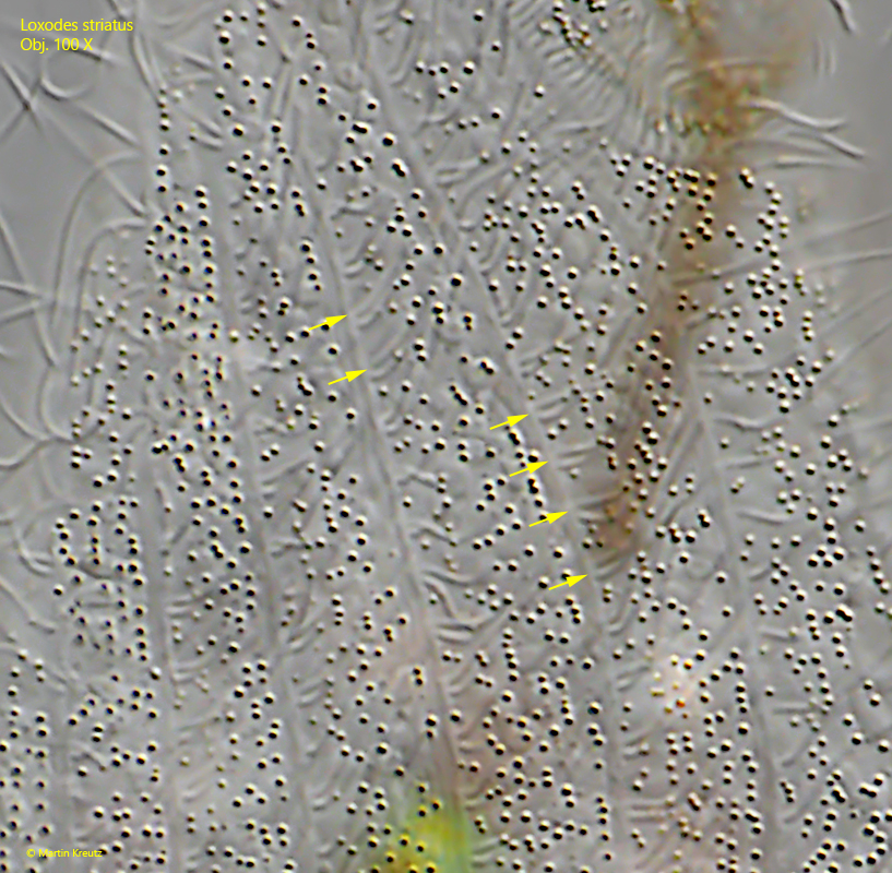
right side with 9–13 longitudinal rows of paired cilia
left side with only two marginal rows of cilia
oral apparatus immediately behind the beak-shaped anterior end
Loxodes striatus
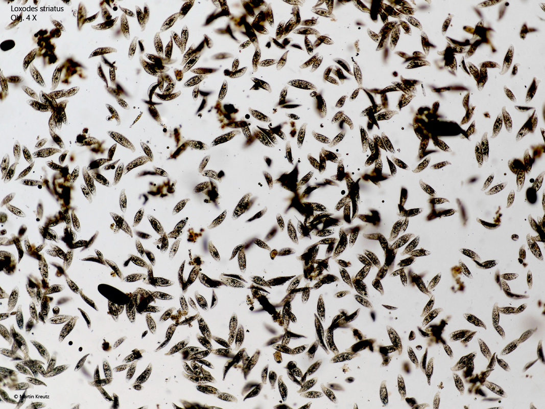
Loxodes striatus is one of the most common ciliates in my samples. It is found in all my sampling sites with a layer of mud and decaying leaves. It often occurs in very large numbers and sometimes there are mass developments where Loxodes striatus is the dominant species in the samples (s. fig. 1).
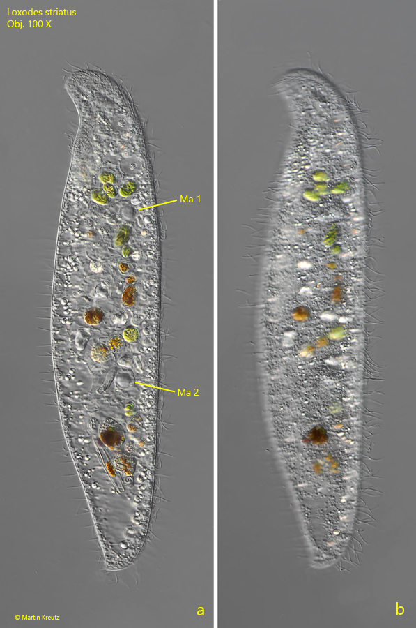
Often Loxodes striatus is found together with the two other species Loxodes rostrum and Loxodes magnus. From the body shape, the three species look similar, due to the curved, beak-shaped anterior end. However, these species can be confidently distinguished by their nuclear apparatus. In Loxodes striatus, two widely separated, spherical macronuclei are present, each with a densely attached micronucleus (s. figs. 4 b and 5 a). Thereby the micronuclei face each other. In Loxodes rostrum the two macronuclei lie close together with one micronucleus between and in Loxodes magnus many macronuclei are distributed in the cytoplasm.
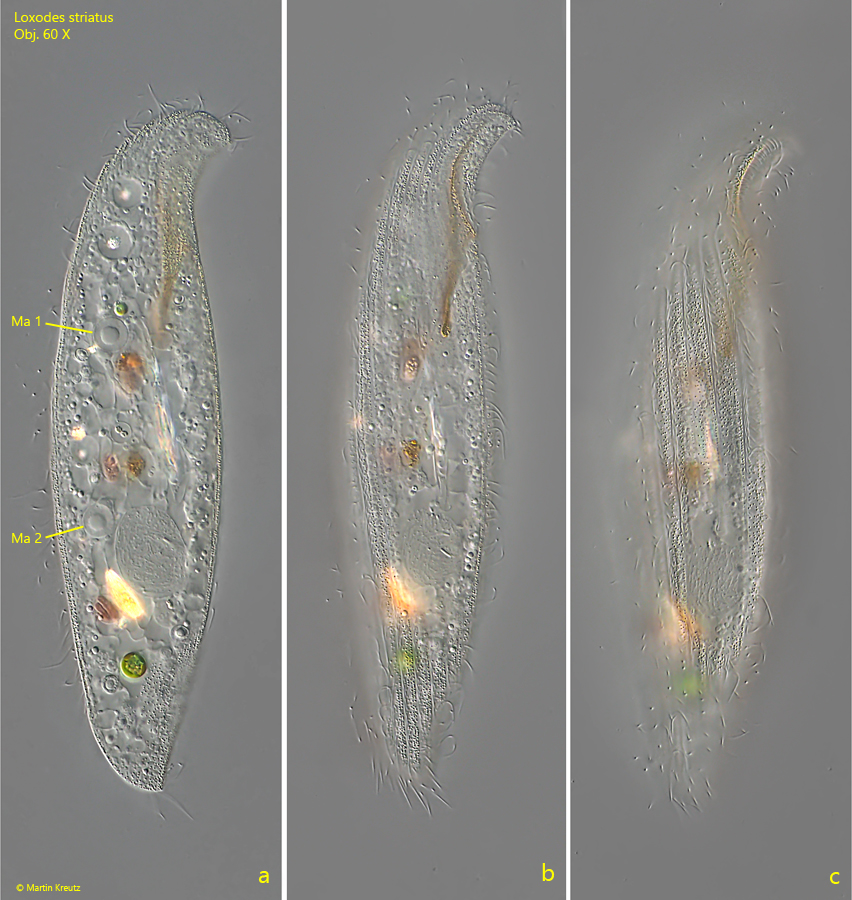
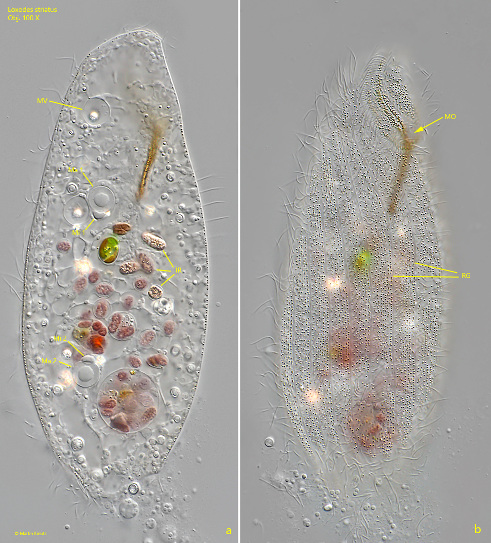
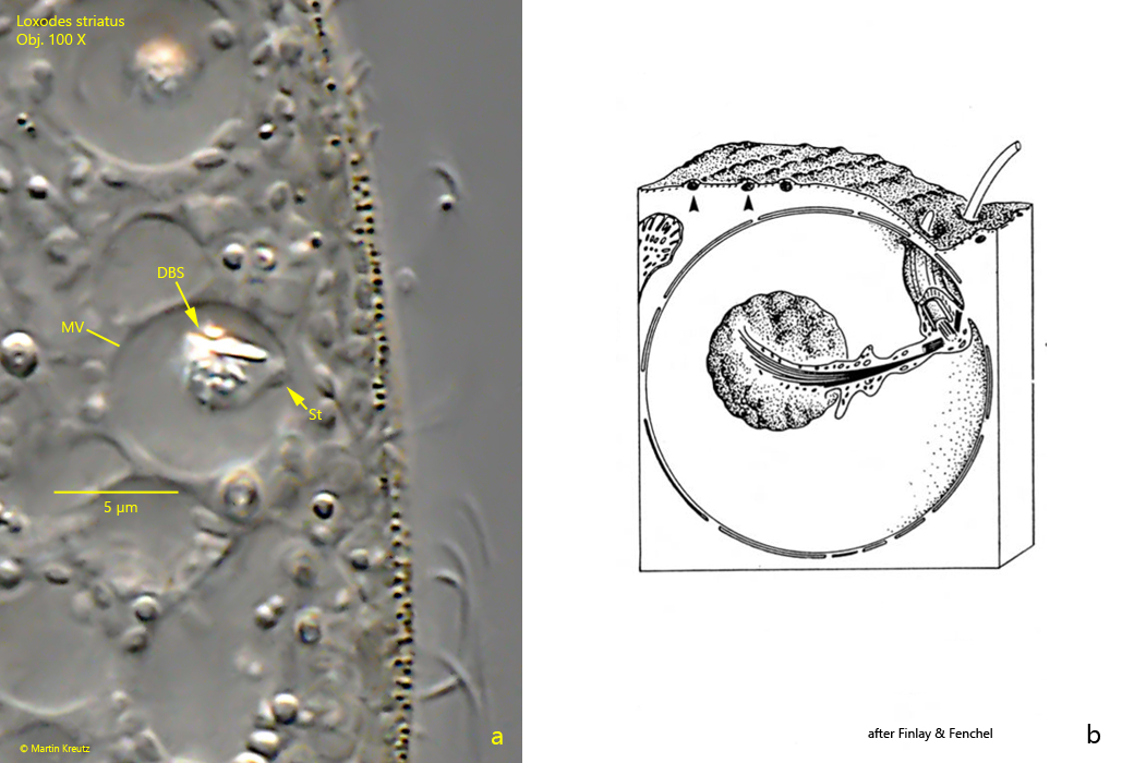
The members of the genus Loxodes have specially constructed Müller vesicles arranged in a row at the dorsal side (s. fig. 4 a). These are constant vacuoles, in which a druse of barium sulfate crystals sits on a cytoplasm stalk (s. fig. 6). It is assumed that this organelle serves for orientation in the water body, similar to an organ of balance. Loxodes striatus has 4–12 of these organelles. They are easily recognized by the highly refractive barium sulfate crystals that shine brightly in DIC (s. figs. 4a and 5a). As another peculiarity, Loxodes has no contractile vacuole (Patterson, 1979), which I find very remarkable. To my knowledge, the way Loxodes striatus regulates its water balance is not known so far.
Fig. 1:Loxodes striatus. A mass development in the Simmelried in September 2022. Obj. 4 X.
Fig. 2 a-b:Loxodes striatus. L = 115 µm. Two focal planes of a freely swimming specimen from left. Ma 1, Ma 2 = macronuclei. Obj. 100 X.
Fig. 3 a-c:Loxodes striatus. L = 171 µm. Three focal planes of a second, freely swimming specimen from right. Ma 1, Ma 2 = macronuclei. Obj. 100 X.
Fig. 4 a-b:Loxodes striatus. L = 202 µm. Two focal planes of a slightly squashed specimen. Note the Müller vesicles (MV) at the dorsal side. Ma 1, Ma 2 = macronuclei, Mi 1, Mi 2 = micronuclei, MO = mouth opening. Obj. 100 X.
Fig. 5 a-b:Loxodes striatus. Two focal planes of a strongly squashed specimen. IR = ingested rhodobacteria, Ma 1, Ma 2 = macronuclei, Mi 1, Mi 2 = micronuclei, MO = mouth opening, MV = Müller vesicles, RG = rows of brownish granula. Obj. 100 X.
Fig. 6 a-b:Loxodes striatus. A Müller vesicle (MV) in detail (a) compared to a schematic drawing. DBS = druse of barium sulfate crystals, St = cytoplasm stalk. Obj. 100 X.
Fig. 7:Loxodes striatus. A crop of fig. 5 b to illustrate the paired cilia, in the rows of cilia on the right side (arrows). Obj. 100 X.