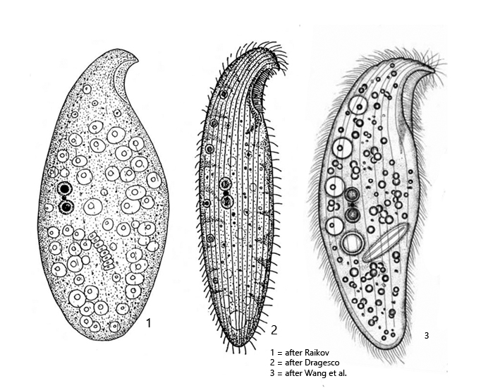
body slender to broadly ellipsoid, anterior end beak-shaped, laterally strongly flattened
length 70–300 µm, width 30–100 µm
two macronuclei close together with a micronucleus between them
no contractile vacuole
a row of about 2–6 Müller vesicles along the dorsal side
cytoplasm filled with ellipsoid and oval shaped symbiotic algae
pellicle with parallel rows of brownish granules (sometimes colorless)
right side with 10–15 longitudinal rows of paired cilia
left side with only two marginal rows of cilia
oral apparatus immediately behind the beak-shaped anterior end
Loxodes rostrum
I find Loxodes rostrum in my samples very often. Often it is associated with the other two species Loxodes striatus and Loxodes magnus. In body shape, these species look similar, due to the curved, beak-shaped anterior end. However, Loxodes rostrum is green in color due to the numerous oval and spindle-shaped symbiotic algae in its cytoplasm. In addition, these species can be distinguished by their nuclear apparatuses. In Loxodes rostrum the two macronuclei lie close together with a micronucleus between them, while in Loxodes striatus they are widely separated with closely apposed micronuclei facing each other. In Loxodes magnus, many macronuclei are distributed in the cytoplasm.
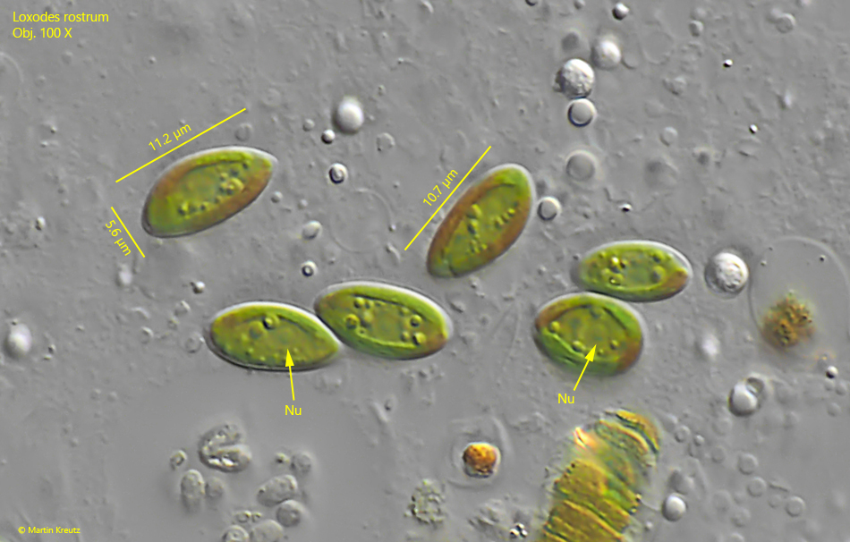
The symbiotic algae of Loxodes rostrum are particularly interesting because they are not of the widely distributed Chlorella type, but are oval or spindle shaped (s. figs. 5 and 6). They are also quite large. According to my measurements, they are 10–12 µm long and 4–6 µm wide (s. fig. 6). I could observe a bell-shaped chloroplast in the algae, which fills almost the entire cell. In addition, each algal cell has its own nucleus (s. fig. 6). To my knowledge, these algae have never been studied in detail and their taxonomic classification and origin is unknown.
The genus Loxodes has specially constructed Müller vesicles. These are constant vacuoles, in which a druse of barium sulfate crystals sits on a plasma stalk. It is assumed that this organelle serves for orientation in the water body, similar to an organ of balance. Loxodes rostrum has 2–6 of these organelles, but they are very difficult to see because of the symbiotic algae.
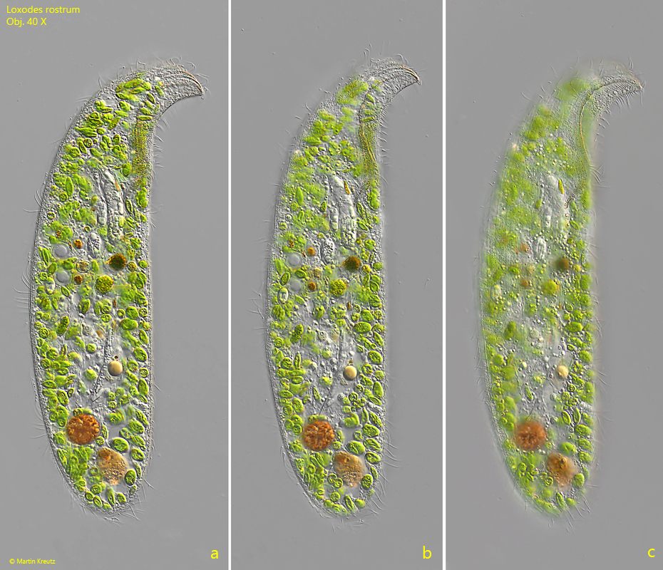
Fig. 1 a-c:Loxodes rostrum. L = 297 µm. Three focal planes of a freely swimming specimen. Obj. 40 X.
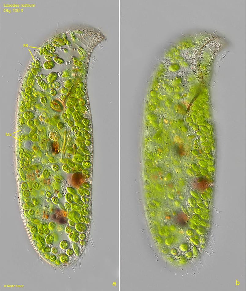
Fig. 2 a-b:Loxodes rostrum. L = 232 µm. Two focal planes of a second, freely swimming specimen. Note the two macronuclei (Ma) close together. SA = symbiotic algae. Obj. 60 X.
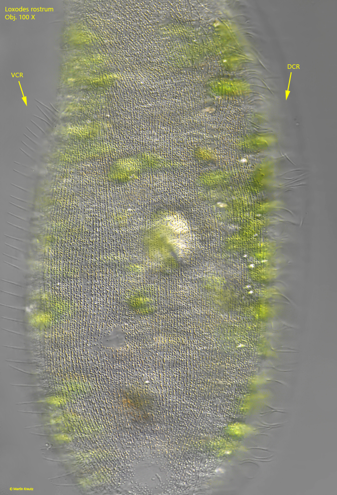
Fig. 3:Loxodes rostrum. The yellow-brownish granula in the pellicle of the left side. The left side is naked apart from from a ventral row of cilia (VCR) and a dorsal row of cilia (DCR). Obj. 100 X.
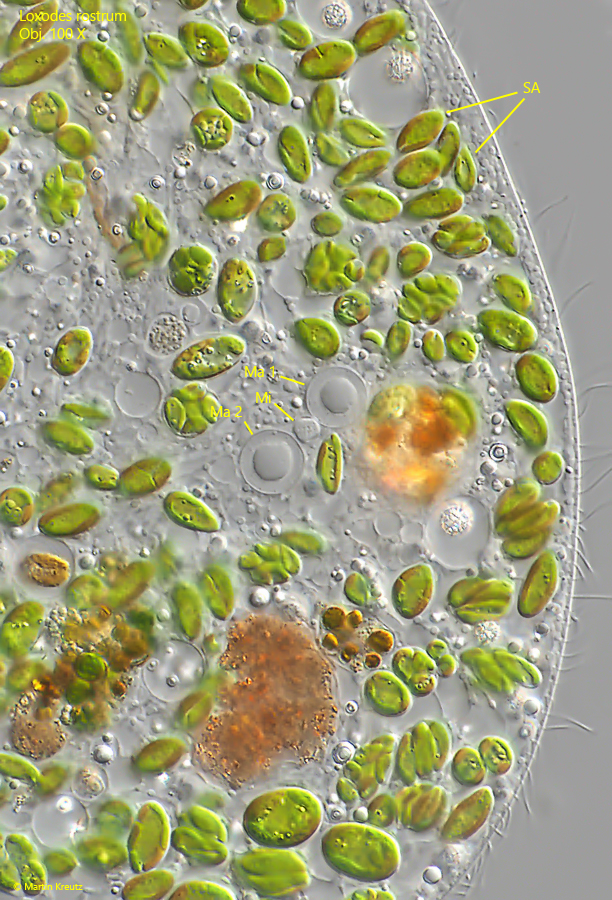
Fig. 4:Loxodes rostrum. The arrangement of the two macronuclei (Ma 1, Ma 2) with the single micronucleus (Mi) in between. Obj. 100 X.
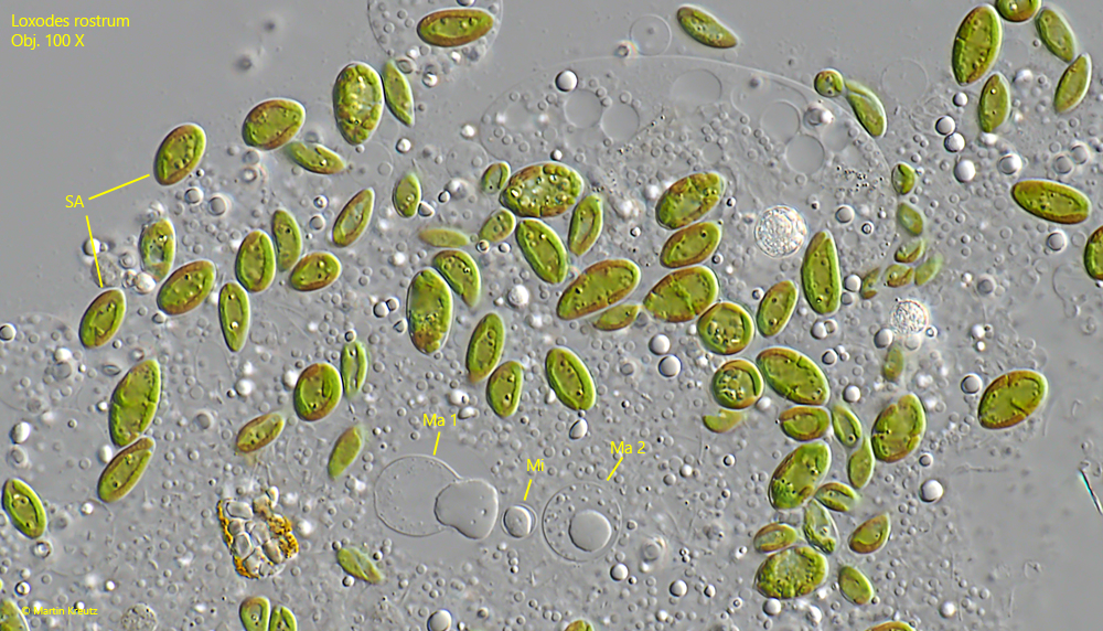
Fig. 5:Loxodes rostrum. Overview of the oval and spindle shaped symbiotic algae (SA) in a strongly squashed specimen. Ma 1 , Ma 2 = macronuclei, Mi = micronucleus. Obj. 100 X.
Fig. 6:Loxodes rostrum. The symbiotic algae in detail. The chloroplast of the symbiotic algae is bell-shaped and fills almost the whole cell. Each algal cell contains a nucleus (Nu). Obj. 100 X.