body oblong with a distinct overhanging apical dome on left side
length 30–40 µm
adoral zone short, 5–6 membranelles, running obliquely before being bent to posterior end
mouth opening near cell equator
perizonal stripe consisting of 5 ciliary rows
one spherical to slightly ellipsoidal macronucleus, diameter 7–8 µm, with many nucleoli
one spherical micronucleus, diameter 2–3 µm
very long caudal cilia (30–50 µm) arise from posterior third
contractile vacuole terminal
Metopus minor
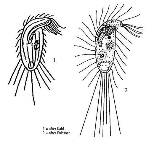
Metopus minor was first described in 1927 by Kahl as Metopus setosus var. minor. His description is very short (2 lines) and is very superficial. In 1980 Foissner published a detailed redescription in which he united the species and varieties Metopus setosus var. minor, Metopus recurvatus and Metopus recurvatus var. pusillus under Metopus minor.
So far I could detect Metopus minor exclusively in the Simmelried. Possibly I have overlooked this species in my other localities because of the small size of Metopus minor. The ciliate is a fast swimmer and rotates around its longitudinal axis. In addition, Metopus minor is very coverslip sensitive, which makes it much more difficult to examine. The species is very characteristic by the protruding apical dome and the long caudal cilia. I could observe that the posterior cell pole is cilia-free and that the caudal cilia arise around this bare field (s. figs. 1 e, 1 c and 2 i). This arrangement was not mentioned by Kahl or Foissner. In some specimens I could see attached bacteria on the pellicle (s. fig. 2 f). I could also observe this phenomenon in Metopus gibbus and Ludio parvulus. Whether these attached bacteria are symbionts or if the ciliate is nerely used as a transport vehicle cannot be decided without further investigation.
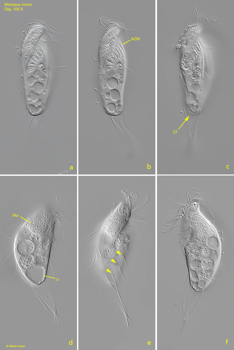
Fig. 1 a-f:Metopus minor. L = 40 µm. A freely swimming specimen from ventral (a-c), right (d, e) and from left (f). Note the cilia free posterior cell pole (CF) and that the caudal cilia arise from the posterior third (arrowheads). The caudal cilia of this specimen are 25 µm long. The adoral zone of membranelles reaches almost the cell equator. CV = contractile vacuole, Ma = macronucleus. Obj. 100 X.
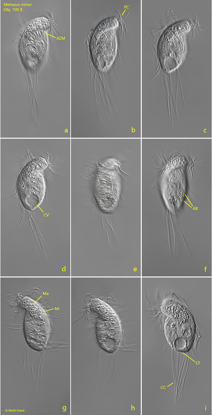
Fig. 2 a-i:Metopus minor. L = 31 µm. A smaller and stockier specimen from ventral (a-f) and from dorsal (g-i). The ventral side of this specimen is covered with adhering bacteria (AB). AZM = adoral zone of membranelles, CC = caudal cilia, CF = cilia free posterior cell pole, Ma = macronucleus, Mi = micronucleus, PC = perizonal cilia. Obj. 100 X.